Wolff-Parkinson-White syndrome (WPW)
About the disease
WPW syndrome is a cluster of symptoms indicating signs of tachycardia on an electrocardiogram (ECG). The name is derived from the first letters of the surnames of the doctors who first described the syndrome: Wolf, Parkinson, and White.
In WPW syndrome, heart palpitations are caused by an atrial-ventricular junction that should not be present in a normal heartbeat. The path of the electrical impulse has a specific route, but an abnormal bundle directly connecting the atrium and ventricle interferes with the normal operation of the conduction system.
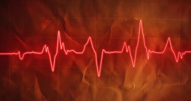
At the pathologic atrial-ventricular junction, the electrical impulse propagates too quickly, which provokes earlier ventricular excitation and is defined as a delta wave on the cardiogram.
The incidence of WPW syndrome is about 0.25% and is more common in males in young adults.
Types
There are four types of WPW syndrome on the ECG.
- Manifesting. Identified by a combination of delta waves and tachycardia on ECG.
- Latent. The patient was diagnosed without signs of delta activity on the cardiogram but with palpitations.
- Multiple. Examination reveals two or more abnormal atrial-ventricular junctions.
- Intermittent. Signs of WPW syndrome are detected intermittently but are not present on the ECG permanently.
If the electrocardiogram shows a characteristic delta wave, but the patient has no signs of arrhythmia, WPW syndrome is not diagnosed. In this case, we are talking about the WPW phenomenon.
Symptoms of WPW syndrome
WPW syndrome is characterized by:
- Regular episodes of palpitations reaching and exceeding 160 wpm;
- dizziness, fainting;
- shortness of breath.
As a rule, clinical manifestations of the disease are not constantly present but occur episodically. Usually, patients complain of a sudden onset of rapid heartbeat, not related to a reaction to external factors (fear, sports), which also stops suddenly. The attacks may last a few minutes or hours.
Reasons
The heart goes through several stages of development during fetal development. By the time the heart is born, the atria and ventricles are isolated from each other, and electrical impulses travel along a predetermined path through the atrioventricular node.
In developmental anomalies in the heart, unnecessary muscle bundles capable of conducting electrical impulses are preserved and not transformed into fibrous tissue. It leads to heart rhythm disturbance – tachycardia, which can be seen on the ECG as a delta wave, an element of blockade of the right leg of the Gis bundle.
Diagnosis
The primary method of confirming the Wolff-Parkinson-White syndrome is an electrocardiogram, which reveals characteristics for the presence of pathological foci of electrical activity in myocardial areas.
Additionally, diagnostics are prescribed to identify the cause of WPW syndrome and assess overall cardiovascular health.
- Echo-CG. Appointed to exclude developmental anomalies and malformations of the heart muscle.
- EFI. The study is necessary to detect the presence of an additional atrial-ventricular junction and determine the features and participation in tachycardia.
- Holter ECG monitoring. Changes on the cardiogram cannot confirm WPW, as the syndrome is a complex of symptoms. Confirming that the patient has regular tachycardia, regardless of workload and emotional state, is essential.
Diagnosis of Wolff-Parkinson-White syndrome may include other tests necessary to rule out other diseases with a similar course and to prepare for surgical treatment.
Treatment
The therapy plan depends on the cause and clinical disease, the severity of the condition, the presence of other serious illnesses, and the drug therapy received for the concomitant pathology.
Conservative treatment
Drug therapy is aimed at normalizing the heart rhythm. The cardiologist prescribes medications for continuous use and for the rapid relief of attacks. In a third of patients, a properly selected complex of drugs protects against attacks for at least one year.
The downside of drug treatment for Wolff-Parkinson-White syndrome is insensitivity to heart rhythm stabilizing drugs, which develops in 50-70% of patients over five years.
Surgical treatment
Radiofrequency catheter ablation is an effective and guaranteed safe treatment method for arrhythmias. During the procedure, the destruction of tissues that trigger an abnormal heartbeat rhythm occurs. That is, during the impact on the myocardium, the foci of pathological electrical activity are destroyed. In WPW, heart syndrome treatment has a higher efficiency than in other causes of tachycardia, so it can be used even without detecting low efficacy and side effects from drugs.
Before the procedure, standard diagnostics are prescribed to assess health and exclude contraindications. Ablation is performed on an empty stomach: 8-10 hours beforehand, it is necessary to refuse food and water. If it is necessary to take any medication, it is necessary to consult a doctor.
Ablation in treating WPW syndrome is performed in a hospital under local anesthesia or anesthesia. A catheter is inserted through the femoral artery or subclavian vein; then, arrhythmogenic areas are identified using cardiac EFI and mapping (recording of the heart’s electrical activity). Current discharges are delivered to destroy the foci of abnormal electrical activity. Low temperatures can be an alternative to current – cryoablation works similarly.
The duration of cardiac ablation averages 2 to 5 hours.
All these treatment options are available in more than 473 hospitals worldwide (https://doctor.global/results/diseases/wolff-parkinson-white-wpw-syndrome). For example, radiofrequency ablation (RFA) can be performed in 23 clinics across Turkey for an approximate price of $6.8 K(https://doctor.global/results/asia/turkey/all-cities/all-specializations/procedures/radiofrequency-ablation-rfa).
Rehabilitation
Patients remain in the hospital under medical supervision for the first day after the ablation. Then, they return home and continue treatment as an outpatient. During 7-10 days after the procedure, the patient may be bothered by moderate chest pain, which is normal. To reduce the load on the heart during the recovery period, limiting physical activity and excluding stress is necessary.
After the rehabilitation period is over, you should follow standard guidelines to maintain a healthy heart:
- controlling salt intake;
- regular exercise;
- no smoking, no alcohol;
- balanced diet;
- normalization of body weight.
Repeat examinations are usually not required.


