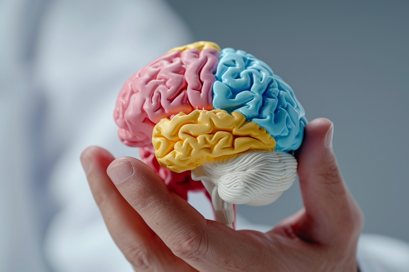
Arteriovenous malformations (AVMs)
About the disease
An arteriovenous malformation (AVM) is a pathologic structure consisting of altered arteries and veins that lack complete capillaries between them. As a result, the tissues around the formation do not receive an entire blood supply. It leads to a violation of their function. In addition, due to pathological changes in the walls of the vessels and the high rate of blood flow in them, there is a high risk of rupture of the malformation and subsequent hemorrhage into the tissues. AVMs can be formed in any part of the body, but most often, doctors find them in the brain or spinal cord. It is this localization that is the most dangerous for the patient.
An arteriovenous malformation is a tangle of twisted vessels. They have many branches that intertwine to form a single conglomerate. It is a relatively rare pathology: brain malformations are found, on average, in one person out of 50 thousand. Most often, the first symptoms of pathology appear relatively early: after 20 years of age. AVMs are diagnosed less frequently in older adults.
The probability of malformation rupture varies between 2-4%. In case of cerebral hemorrhage, every tenth patient dies, and half of the people become disabled. The only way to get rid of the pathology is surgery.
Classification of arteriovenous malformations
There are several types of classification of arteriovenous malformations, each of which is important in the choice of treatment tactics. Depending on the localization, there are formations of the brain, spinal cord, skin, etc.
Features of the structure of the formation allow us to distinguish three forms of AVMs:
- Angiomatous: a network of intertwined vessels;
- Fistulous: the artery passes directly into a vein without forming a vascular tangle;
- Mixed: combines angiomatous and fistulizing forms.
Brain malformations are also categorized based on size:
- small: diameter less than 3 cm;
- medium: 3-6 cm in diameter;
- large: size 6 centimeters or more.
Neurologists and neurosurgeons use a scoring system to assess the degree of surgical risk for cerebral AVMs. They consider the size, hemodynamic features, and location of the mass. It is necessary for the choice of tactics for treating the mass.
Symptoms of arteriovenous malformations
Signs of AVMs depend on their size and location. If the size is small, the pathology is asymptomatic and is often an incidental finding. Symptoms of cerebral arteriovenous malformations often resemble the signs of a tumor. The patient may be bothered by the following:
- headaches;
- dizzy spells;
- visual or hearing impairment;
- wobbly walking;
- movement coordination disorders;
- seizures;
- episodes of unconsciousness;
- decreased muscle tone, etc.
When AVMs develop in the spinal cord, patients complain of back pain, internal organ dysfunction (e.g., urinary incontinence), sensory or muscle tone disorders in the extremities, etc.
Cutaneous malformations are accompanied by edema or hypertrophy of tissue, ulceration of the skin, gangrene, and severe pain in the area of the lesion.
When a central nervous system AVM ruptures, the patient shows signs of hemorrhagic stroke or spinal cord hemorrhage. These are severe pain in the head and back, signs of irritation of the brain membranes, loss of consciousness, and sudden paralysis. The condition is life-threatening and requires immediate medical attention.
Causes of arteriovenous malformations
The exact causes of AVM development are currently unknown. Most of the detected anomalies are congenital and are formed at the stage of intrauterine development. It is believed that the risk increases against the background of toxic effects on the fetus (taking certain drugs by a pregnant woman, smoking, alcoholism, drug addiction), the influence of ionizing radiation, and a number of diseases (infections, diabetes mellitus, bronchial asthma, and others). Acquired forms of AVMs, which are formed as a result of tissue trauma, are rare.
Diagnosis of arteriovenous malformations
Neurologists, orthopedists, or therapists can perform the initial examination of patients with malformations, and then vascular surgeons and neurosurgeons join them. In the first stage, complaints and anamnesis are collected, and the dynamics of the development of each symptom and the characteristics of the patient’s lifestyle are assessed. Then, the person is referred for instrumental examination.
- Ultrasound and duplex scanning of vessels. It allows the detection of the pathological focus, assesses the speed and direction of blood flow, and distinguishes AVMs from other volumetric masses (tumors).
- Angiography. The injection of a contrast agent into the blood vessels allows them to be mapped on a series of X-rays and visualize the AVM.
- CT and MRI. They are performed when other methods are not sufficiently informative and require contrasting vessels.
Additionally, the patient may be prescribed laboratory diagnostics, EEG, consultations with specialized specialists of related profiles, and other examinations.
Treatment of arteriovenous malformations
Malformations are treated only surgically, as there are no conservative ways to restore the structure of the altered arterial and venous vessels. Depending on the situation, surgeons may use different types of interventions.
- AVM resection. Ligation of the feeding vessel with subsequent removal of the malformation and surrounding tissues. As a rule, the intervention is performed openly.
- Endovascular occlusion (embolization) and sclerotherapy. Special substances that block blood flow are injected into the vessel feeding the AVM. Without a blood supply, the formation gradually undergoes reverse development.
- Stereotactic radiosurgery. High-precision irradiation of the AVM area causing coagulation of the vessels.
Surgical interventions are always risky for the patient, which is why doctors recommend that they be performed routinely after thorough preliminary examination and preparation. Emergency surgery is only performed in life-threatening situations, for example, in cases of ongoing bleeding.
All these treatment options are available in more than 490 hospitals worldwide (https://doctor.global/results/diseases/arteriovenous-malformations-avms). For example, arteriovenous malformations (AVM) embolization can be performed in 8 clinics across Turkey for an approximate price of $8.5K(https://doctor.global/results/asia/turkey/all-cities/all-specializations/procedures/arteriovenous-malformations-avm-embolization).
Rehabilitation
The duration of rehabilitation after AVM removal depends on the size and localization of the pathological formation, the type of intervention, and the patient’s initial condition. The period of hospitalization varies from 1-2 days to several weeks. After discharge, a person should avoid physical activity, lifting weights, heat procedures, and exposure to ultraviolet rays. It is essential to keep blood pressure levels under control and take medications prescribed by the doctor, including those to prevent thrombosis. Restrictions last 1-3 months. It is necessary to see a doctor and undergo diagnostics periodically during this period.