Brain cyst
What is a brain cyst?
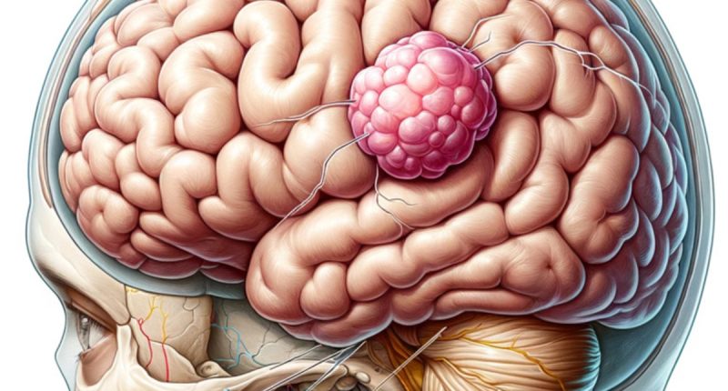
A brain cyst is a hollow formation consisting of membranes or locally compressed brain tissue filled with fluid. The cyst is often filled with cerebrospinal fluid; in some cases, its contents are products of microorganisms. Cysts rarely accompany the development of a tumor process in the brain.
Often, the cyst develops slowly and progresses asymptomatically. However, in case of significant size, compression of brain tissues is possible with the appearance of focal syndrome, the manifestations of which depend on the localization of the formation.
Cysts can be congenital or acquired. They are found in patients of any age (from newborns to the elderly). Cysts can be single or multichambered. They are classified according to their nature, localization, and origin (arachnoid, pineal, vascular, colloid, dermoid, tumor).
Symptoms of a brain cyst
Most congenital cysts do not manifest themselves during life and do not pose an oncologic risk. Large acquired masses cause symptoms of high intracranial pressure, namely:
- headaches;
- decreased ability to work;
- trouble sleeping;
- a feeling of pressure in the eyeball area;
- visual hallucinations;
- focal seizures (face, limbs);
- pulsation in the head;
- nausea and vomiting unrelated to food intake.
In children, psychomotor development may be impaired. In some cases, cyst formation carries the risk of cerebral hemorrhage, which can lead to severe consequences (disability, death).
Causes of brain cysts
Congenital brain cysts are formed in case of an unfavorable impact on the fetus at the antenatal stage. The provoking factors may be intrauterine hypoxia, fetoplacental insufficiency, Rhesus conflict, infectious complications, the mother’s intake of drugs with teratogenic properties, chronic intoxication in drug addiction, alcoholism, or tobacco smoking.
The formation of an acquired cyst can be triggered by:
- trauma to the newborn’s head during labor;
- brain injuries at any age;
- inflammatory diseases of the brain;
- cerebral circulatory disorders;
- parasitic infections;
- post-operative complications.
Neuroinfections, repeated head trauma, vascular disorders, inflammatory processes, and other factors can cause a growth spurt of an existing intracerebral mass.
Diagnosis of brain cysts
Diagnosing a brain cyst typically involves:
- Neurological Examination: Assessing brain function through various tests.
- Imaging Tests: MRI or CT scans are used to get detailed images of the brain.
Suspicion of a volumetric mass in the brain arises during evaluating the patient’s complaints and neurological status. Increased intracranial pressure is confirmed by ultrasound; in seizures and epileptic paroxysms, electroencephalography is performed.
However, even with careful examination of all this information, it is impossible to distinguish a cyst from a brain tumor or abscess. CT or MRI allows visualization of the formation. To distinguish a cyst from a tumor, studies with contrast are performed because the latter do not accumulate in them. It is important not only to detect the mass but also to monitor its dynamic changes.
Treatment of brain cysts
Treatment for brain cysts varies:
- Observation: Small, asymptomatic cysts may only require regular monitoring;
- Medication: To manage symptoms like seizures or headaches;
- Surgical Intervention: This may be necessary for larger cysts causing significant symptoms. Techniques include;
- Drainage: Draining fluid from the cyst;
- Excision: Surgical removal of the cyst.
Conservative approach in the treatment of brain cysts is ineffective. Dynamic observation is indicated for congenital formations of small size. Surgery is prescribed for patients in whom the cystic cavity increases and there is a risk of complications (rupture, hemorrhage, etc.).
Urgent operations with skull trepanation are performed on patients with severe disorders caused by compression of brain tissue and high intracranial pressure.
Cyst removal surgery is usually performed routinely using endoscopic equipment. The intervention involves draining the cyst through a small milled hole in the skull. After surgery, the patient undergoes rehabilitation, which involves neuropsychologists, physical therapy, and reflexology.
Cyst removal surgery is performed in more than 50 hospitals worldwide (https://doctor.global/results/diseases/brain-cyst). For example, Brain cyst surgical treatment is done in 11 clinics across Turkey for an approximate price of $6.8 K(https://doctor.global/results/asia/turkey/all-cities/all-specializations/procedures/brain-cyst-surgical-treatment).
Rehabilitation
Rehabilitation post-treatment may involve:
- Physical therapy to regain balance and coordination.
- Occupational therapy for daily living skills.
- Cognitive therapy for memory, attention, or other cognitive issues.
Outcomes
The outlook for individuals with brain cysts varies:
- Many live symptom-free lives, especially with smaller cysts.
- Surgical removal can often lead to a good prognosis, though risks are involved.
- Regular follow-up is crucial to monitor for any changes or recurrence.
Conclusion
Brain cysts are a diverse group of conditions that can range from asymptomatic to causing significant health issues. Understanding the causes and symptoms is vital for early detection and treatment. While many cysts are benign and may require only monitoring, others necessitate medical intervention or even surgery. The outcomes for patients with brain cysts are generally favorable, especially with early diagnosis and proper management. As with any medical condition, it’s important for individuals with brain cysts or those experiencing related symptoms to consult healthcare professionals for accurate diagnosis and appropriate treatment plans. Regular follow-ups and adherence to treatment and rehabilitation programs play a key role in managing the condition and maintaining quality of life.

