Ankle fracture
What’s that?
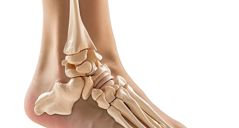
An ankle fracture is a disruption of the integrity of the fibula and/or tibia in the distal segment involved in forming the ankle joint.
About the disease
Ankle fractures account for approximately 10-20% of leg fractures. These injuries are most often associated with an outward or inward turning of the leg. In about one in three cases, ankle fractures are combined with a lower interosseous membrane, which is also referred to as the middle tibiofibular ligament.
The severity of the injury determines clinical symptoms. Complete fractures are more severe than incomplete fractures. The final diagnosis is based on the results of radiologic scanning.
The stability of the injury determines the treatment of ankle fractures. In stable fractures, there is virtually no risk of displacement of bone fragments, so conservative tactics are applicable. In unstable fractures, there is an increased likelihood of displacement of bone fragments, so additional fixation is required. For this purpose, surgical intervention is performed. In addition, after surgery, recovery is faster, and a person returns to his usual way of life faster (most often two months after the injury).
Types of ankle fractures
According to the classification, the following types of ankle fractures are distinguished:
- injuries with displaced bone fragments and injuries without displaced bone fragments;
- injuries with the damage to the integrity of the bone base of the outer ankle, inner ankle, or combined injuries where both ankles are injured simultaneously.
Most ankle fractures are repositionable and stable after immobilization with a leg cast. Unstable injuries include those with external foot subluxation or disruption of two of the three stability complexes (i.e., more than one ankle or a combination of damage to the external ankle and intercostal syndesmosis).
Symptoms of an ankle fracture
Symptoms of an ankle fracture may include the following:
- painful sensations in the ankle joint associated with an injury;
- gradual onset of soft tissue edema;
- the lividity of the skin associated with hemorrhage into the soft tissues;
- impaired leg function – incomplete fractures may impair support and movement, while complete fractures make it impossible to step on the injured leg;
- increased pain when feeling the ankle area;
- presence of pain in the middle third of the tibia with moderate compression of the ankles in the direction of each other.
In a complete ankle fracture, the bone fragments assume an atypical position, resulting in a shin deformity.
Causes of ankle fracture
The average age of the patients was 45 years, with a predominance of postmenopausal women and younger male patients. These are low-energy injuries due to a fall from a height less than one’s height, caused in most cases by forced external rotation and supination or pronation and abduction of the foot. An ankle fracture can also be caused by direct impact with a heavy object on the lower leg.
Diagnosis
According to clinical guidelines, diagnosing an ankle fracture of the tibia includes an objective examination and X-ray scan.
Radiologic signs indicating disruption of bone integrity:
- visualization of the lesion line, which appears dark in comparison to the bone tissue;
- atypical positioning of bone segments;
- the presence of displaced bone fragments;
- displacement of bones outside the ankle joint (in the presence of concomitant dislocation).
In complex clinical cases, a CT or MRI scan is indicated. These methods allow a detailed assessment of the ankle joint and careful planning of the upcoming surgery.
Treatment of an ankle fracture
Both conservative and surgical methods can carry out treatment of ankle fractures. The nature of the injury determines the choice of treatment program.
Conservative treatment
Cast immobilization is used both in stable ankle fractures as the primary treatment method and as an additional method after immersion osteosynthesis of unstable fractures and does not significantly affect the long-term results. Conservative treatment tactics can also be applied if the patient has contraindications to surgery; in particular, it may be a severe concomitant pathology, which increases the anesthetic risks.
Conservative management involves manual repositioning of the bone fragments and the application of an immobilizing dressing. It can be made of plaster or polymer. The dressing length is from the heads of the metatarsal bones to the top of the tibia.
Soon after the immobilizing dressing is applied, the patient is allowed to walk on crutches but avoid supporting the injured leg. About one month later, a control X-ray scan is performed. If, according to the X-ray data, a bone callus has formed, dosed support on the injured leg is allowed. The weight of your own body can be transferred to the foot in 1.5-2 months after the injury if the radiography results are satisfactory. A repeat radiologic evaluation is performed two months after the injury. If the bone tissue has regenerated, the immobilizing dressing is removed, and a period of active development of the limb begins.
Surgical treatment
Surgical intervention is performed for unstable fractures, including foot dislocation. The surgery is performed under spinal anesthesia. It consists of the fixation of the bone fragments with the help of metal structures. Today, there are methods of internal fixation of ankle fractures, which consist of open repositioning and osteosynthesis of fractures of the inner and outer ankle, the posterior edge of the distal metaepiphysis of the tibia with tightening screws, wire loops, and tip plates.
Open surgery conventionally consists of 4 stages:
- The first stage is the open reduction of the fracture and manual bone repositioning in the presence of foot dislocation. First, the anatomy of the external ankle is restored, after which the fragments are fixed with a screw in a direction perpendicular to the fracture line. Additionally, a metal plate is also applied.
- The second stage is to restore the structure of the inner ankle. Usually, two screws are used to prevent rotation of the bone fragments.
- The third stage assesses the preservation of the interosseus connective tissue. If a tear is diagnosed, it is fixed with a special technique.
- The fourth stage is layer-by-layer tissue repair of the surgical area. A drainage tube may be left in the wound to access the external ankle.
An alternative functional surgical method for unstable ankle fractures is closed percutaneous osteosynthesis. This technique is performed using the Ilizarov apparatus.
All these treatment options are available in more then 750 hospitals worldwide (https://doctor.global/results/diseases/ankle-fracture). For example, Ankle fraction surgery is performed in 45 clinics across Germany for an approximate price of $10.9 K (https://doctor.global/results/europe/germany/all-cities/all-specializations/procedures/ankle-fracture-surgery).
Prevention
There is no specific prevention for this disease. All ankle fracture prevention is aimed at preventing injury.
Rehabilitation after ankle fracture
Comprehensive and timely rehabilitation after an ankle fracture will prevent possible complications. Thus, among the unfavorable consequences may be ankle contractures, muscle atrophy, deterioration of the foot support function, flat feet, deforming arthrosis, and violation of the correct gait pattern.
What does an ankle fracture mean from a rehabilitologist’s point of view? Massage, therapeutic exercise, and physiotherapy play a significant role in restoring the functionality of the leg. Recently, kinesiotherapy methods have been widely used. Rehabilitation is conditionally divided into three stages:
- Immobilization period. The methods stimulate the regeneration of damaged tissues, prevent muscle atrophy, osteoporosis, and contractures, and prepare the shoulder girdle muscles for walking on crutches. At this stage, therapeutic gymnastics, therapeutic massage, and physiotherapy are performed.
- Postimmobilization period. At this stage, hydrokinesis therapy, therapeutic gymnastics, therapeutic massage, and physiotherapy are recommended. These measures aim to restore the supporting function of the limb, accelerate the processes of bone callus structuration, develop the strength and endurance of muscles of the injured limb, adapt to early dosed loads, and improve the processes of blood circulation and lymphatic outflow.
- Training period. Kinesiotherapy, self-massage, and gymnastics are performed, and wearing orthopedic insoles with a supinator is also recommended. At this stage, the joint and limb functions are fully restored, the risks of deforming arthrosis and flat feet are reduced, the walking and support function is improved, and the leg is prepared for prolonged static and dynamic loads during everyday activities.


