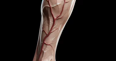
Benign bone tumor
Definition
Osteoma is a benign tumor that develops from bone tissue. It is characterized by a favorable course: it grows very slowly, never maligns, does not metastasize, and does not sprout into surrounding tissues. Usually, osteomas are localized on the outer surface of bones and located on the skull’s flat bones, in the walls of maxillary, lattice, cuneiform, and frontal sinuses, and on the tibia, femur, and humerus. Vertebral bodies may also be affected. Osteomas are usually single; the exception is Gardner’s disease, which is characterized by multiple tumors and congenital osteomas of the bones of the skull due to a violation of mesenchymal tissue development and combined with other malformations. Treatment of all types of osteomas is surgical only.
General information
Osteoma is a benign tumor-like mass formed from highly differentiated bone tissue. It is characterized by extremely slow growth and a very favorable course. No cases of osteoma degeneration into a malignant tumor have been identified. Depending on the variety, osteoma may be accompanied by pain or be asymptomatic. If neighboring anatomical formations (nerves, vessels, etc.) are compressed, there is a corresponding symptomatology that requires surgical intervention. In other cases, surgical removal of osteomas is usually performed for cosmetic reasons.
Osteomas usually develop in childhood and adolescence. Male patients are more often affected (except for osteomas of the facial bones, which develop more often in women). Gardner syndrome, accompanied by the development of multiple osteomas, is hereditary. In other cases, it is assumed that hypothermia or repeated trauma may be the provoking factors.
Classification
Considering the origin, two types of osteomas are distinguished in traumatology:
- Heteroplastic – develop from connective tissue. This group includes osteophytes. They can appear not only on bones but also in other organs and tissues: in places of tendon attachment, the diaphragm, pleura, brain tissue, heart membranes, etc.
- Hyperplastic – develop from bone tissue. This group includes osteomas and osteoid osteomas.
- Osteoma does not differ in its structure from normal bone tissue. It is formed on the bones of the skull and facial bones, including the walls of the sinuses (frontal, maxillary, lattice, cuneiform). Osteoma in cranial bones is two times more common in men, and in facial bones, it is 3 times more common in women. In the vast majority of cases, single osteomas are detected. In Gardner’s disease, forming multiple osteomas in the region of long tubular bones is possible. In addition, there are congenital multiple osteomas of the bones of the skull, which are usually combined with other malformations. The osteomas themselves are painless and asymptomatic, but when squeezed by neighboring anatomical structures, they can cause a variety of clinical symptoms, from visual impairment to epileptic seizures.
- Osteoid osteoma is also a highly differentiated bone tumor, but its structure differs from normal bone tissue and consists of heavily vascularized areas of osteogenic tissue, chaotically arranged bone beams, and areas of osteolysis (bone destruction). Typically, an osteoid osteoma does not exceed 1 cm in diameter. It is standard and accounts for about 12% of all benign bone tumors.
- Osteophytes can be internal or external. Internal osteophytes (enostoses) grow into the medullary canal, are usually solitary, are asymptomatic, and become an incidental finding on radiographs. External osteophytes (exostoses) grow on the surface of the bone. They may develop as a result of various pathologic processes or occur for no apparent reason. Exostoses can be asymptomatic, appear as a cosmetic defect, or compress neighboring organs.
Osteoma
Symptoms
The clinic of an osteoma depends on its location. When an osteoma is localized on the outside of the skull bones, it presents as a painless, immobile, very dense mass with a smooth surface. Osteoma, located on the inner side of the bones of the skull, can cause memory disorders, headaches, increased intracranial pressure, and even become the cause of the development of epileptic seizures. An osteoma localized in the area of the Turkish saddle can cause hormonal disorders.
Osteomas of the long tubular bones are usually asymptomatic and are detected when Gardner’s disease is suspected or becomes an incidental finding in radiologic studies.
Diagnosis
Additional studies are used to diagnose osteoma. Initially, radiography is performed. However, due to osteomas’ small size and peculiar location (for example, on the inner surface of the skull bones), this study is not always effective. Therefore, more informative computed tomography often becomes the primary diagnostic method.
The differential diagnosis of osteomas of the facial and cranial bones is made with solid odontoma, ossified fibrous dysplasia, and reactive bone growths that may occur after severe trauma and infectious lesions. Osteomas of long tubular bones must be differentiated from osteochondroma and organized periosteal callus.
Treatment
Depending on their localization, osteomas are treated by neurosurgeons, maxillofacial surgeons, or traumatologists. Surgery is indicated for cosmetic defects or symptoms of compression of neighboring anatomical structures. If the osteoma is asymptomatic, dynamic observation is possible.
Osteoid osteoma
Characteristics
Osteoid osteoma can be found on any bone except the sternum and skull. Its typical localization is the diaphyses and metaphyses of the long tubular bones of the lower extremities. About half of all osteoid osteomas are found on the tibia and in the proximal metaphysis of the femur. Osteoid osteoma develops at a young age and is more often observed in men. It is accompanied by increasing pain, which appears before the onset of radiologic changes. Vertebral osteoid osteomas account for approximately 10% of the cases.
Symptoms
The first symptom of osteoid osteoma is limited pain in the lesion area, which initially resembles muscle pain. Subsequently, the pain becomes spontaneous and progressive. The pain syndrome in such osteomas decreases or disappears after taking analgesics and after the patient becomes active but reappears at rest.
At the beginning of the disease, no external changes are detected. Then, a flat and thin painful infiltrate forms over the area of the lesion. When osteoid osteoma occurs in the epiphysis region, fluid accumulation may be determined in the joint. When located near the growth zone, osteoid osteoma stimulates bone growth, causing children to develop skeletal asymmetry. If the osteoid osteoma is localized in the vertebral region, scoliosis may form.
Diagnosis
Osteoid osteoma is diagnosed based on a characteristic radiologic picture. Due to their location, these tumors are usually more visible on X-rays than a normal osteoid osteoma. In some cases, however, osteoid osteoma may also be difficult to diagnose due to its small size or localization (e.g., in the vertebral region). In such situations, a CT scan is used to clarify the diagnosis.
Radiologic examination reveals a small rounded area of lumen under the cortical plate surrounded by a zone of osteosclerosis, which increases in width as the disease progresses. At the initial stage, there is a clearly visible border between the rim and the central zone of the osteoma. Later, this boundary is obliterated as the tumor undergoes calcification.
The differential diagnosis of osteoid osteoma is made with limited sclerosing osteomyelitis, dissecting osteochondrosis, osteoperiostitis, chronic Brody’s abscess, and, less commonly, Ewing’s tumor and osteogenic sarcoma.
Treatment
Osteoid osteoma is usually treated by traumatologists and orthopedists. Treatment is surgical only. If possible, the affected area and the surrounding area of osteosclerosis are resected during surgery. Recurrences are very rare.
Osteophytes
Characteristics
These growths can occur for various reasons and differ from classical osteomas in several characteristics. However, due to their similar structure – highly differentiated bone tissue – some authors classify osteophytes as osteomas.
Of practical interest are exostoses – osteophytes on the outer surface of the bone. They can have the shape of a hemisphere, a mushroom, a spike, or even a cauliflower. Hereditary predisposition is noted. Formations more often occur during puberty. The most common exostoses are in the upper third of the tibia, lower third of the femur, upper third of the humerus, and lower third of the forearm bones. Less frequently, exostoses are localized on the flat bones of the trunk, vertebrae, hand, and metatarsal bones.
Diagnosis
The diagnosis is made based on radiography and/or CT scan data. When studying X-ray images, the actual size of the exostosis does not correspond to the data because the upper cartilage layer is not shown on the images. At the same time, the thickness of this layer (especially in children) can reach several centimeters.
Treatment
The treatment is operative, performed in the department of traumatology and orthopedics, and consists of removing the exostosis. The prognosis is good; recurrences in single exostoses are rare.
All these types of treatment are available in more than 520 hospitals worldwide (https://doctor.global/results/diseases/benign-bone-tumor). For example, Surgery for bone cancer can be done in 23 clinics across Turkey for an approximate price of $5.4 K (https://doctor.global/results/asia/turkey/all-cities/all-specializations/procedures/surgery-for-bone-cancer).

