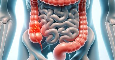
Benign rectal tumor
General information
Benign tumors of the rectum are a local formation associated with the active division of cells of the mucosa or other layers of the organ. Such neoplasms can create a mechanical obstacle to the movement of fecal matter and may also increase cancer risks.
Benign tumors of the rectum have a high degree of differentiation, so their structure is similar to normal cells of the rectal structures. Such neoplasms do not metastasize and do not infiltrate the surrounding tissues, which is their main difference from malignant tumors. However, at a particular stage, mutations may occur in some benign formations that predispose to the development of rectal cancer. It explains the increased attention of proctologists to such tumors.
In addition, benign neoplasms can become large and block the intestinal lumen. Thus, intestinal obstruction develops, often indicating emergency surgery. Therefore, timely diagnosis and treatment are essential.
Types
Benign tumors of the rectum are of 3 types according to their histological structure.
- Adenomas originating from the mucosa’s covering epithelium are the most common. The adenoma may be villous, tubular, or mixed (villous-tubular).
- Adenomatous polyps also originate from glandular cells. Because they pose the most significant cancer risk, all polyps must be sent for morphologic examination after removal.
- Serrated dysplasia is one of the rare benign tumors of the rectum.
Non-epithelial neoplasms may develop in the terminal colon. These are:
- Lipomas – originating from adipose tissue;
- Angiomas – develop from blood vessels;
- Fibromas – the source is connective tissue, etc.
Tumors of any histologic structure may be mushroom-shaped, where there is a stalk, or attached to the intestinal wall by a broad base.
The number of benign neoplasms can be different. Therefore, single and multiple tumors of the rectum are distinguished.
Symptoms
Most benign tumors of the rectum are asymptomatic. These lesions are detected accidentally during endoscopic examination. However, a small proportion of these tumors can malignize, which is the most common cause of colorectal cancer.
Determining morphologic analysis is required to determine the degree of dysplasia and predict the likelihood of malignant transformation. Severe dysplasia combined with a tumor size greater than 1 cm is associated with a high incidence of rectal cancer. These risks increase if the tumor morphology presents predominantly with a villous component. Carcinogenesis is associated with specific mutations in adenomatous cells.
In cases where benign tumors have a clinical manifestation, the main symptoms may be as follows:
- prolapse of the neoplasm from the rectum during defecation;
- mucus secretion (most characteristic of villous adenoma);
- itching, burning, and discomfort in the anal area;
- impurity of blood to fecal masses (its appearance is associated with traumatization of the formation during the passage of fecal masses);
- wetness of the perianal area;
- painful sensations in the rectum are rarely intense, so patients are delayed in seeking medical attention.
Reasons
The main causative factors leading to the development of benign rectal tumors are:
- digestive disorders;
- excessive consumption of red meats (pork, beef, lamb), which have been identified as potential carcinogens;
- errors in the diet associated with the use of smoked foods and convenience foods;
- insufficient proportion of fruits and vegetables in the daily diet (a day in the human body should be 30 g of fiber, necessary for the correct functioning of the digestive tract);
- consumption of refined foods high in saturated fat;
- excessive consumption of alcoholic beverages;
- infections affecting the large intestine;
- hypodynamia;
- overweight and obesity;
- aggravated heredity – the presence of benign and malignant digestive tract and ovary tumors in first and second-line relatives.
Diagnosis
The diagnostic search begins with a detailed anamnesis collection. After this, the proctologist proceeds to the objective stage, which includes a visual assessment of the rectal region and a palpatory examination of the rectum. However, the final diagnosis of neoplasms of the terminal part of the digestive tract is established after endoscopic examination. Under anesthesia, the proctologist introduces an endoscope into the rectum and examines the state of the mucosa in detail. The enlarged image is displayed on the screen.
The advantages of endoscopic examination are apparent. First, if atypical changes are suspected, the proctologist can take a tissue sample and send it for histologic examination. Only the study of morphology under a microscope can accurately determine whether the tumor is benign or malignant.
Second, if there is a certainty that the polyp, adenoma, or other neoplasm does not have atypical changes, it can be removed in one stage. Biomaterial, in its entirety, is sent to a morphologic laboratory. We can talk about radical treatment only after the histologist’s conclusion with certainty.
Conservative treatment
The primary method of treatment of benign tumors is surgical. Such a radical approach is associated with a potential risk of oncologic transformation.
Benign rectal tumors are not treated with medication. Observational and wait-and-see tactics are also not used.
Surgical treatment
Modern methods of treatment of benign rectal tumors are endoscopic loop excision and transanal microsurgery. In the presence of large adenomas, surgeons sometimes have to perform intra-abdominal interventions, which are accompanied by high traumatism, increased incidence of postoperative complications, and violation of the function of the locking apparatus. Removal of benign rectal tumors involves the use of general anesthesia. After anesthesiologists select the drugs individually, the patient falls asleep without feeling any discomfort during the intervention.
With the laparoscopic method, the surgeon makes several small holes in the anterior abdominal cavity through which special instruments and a video camera are inserted. Carbon dioxide is injected into the abdominal cavity to improve the view. Under control of the image on the monitor, the doctor excises the affected part of the rectum, and the healthy ends are stitched together, restoring the typical passage of contents through the digestive tract.
Cavity surgery involves opening the anterior abdominal wall with a scalpel. This variant of intervention is used in cases of large tumor size when its removal is impossible through tiny openings.
All these treatment options are available in more than 700 hospitals worldwide (https://doctor.global/results/diseases/benign-rectal-tumor). For example, Transanal endoscopic microsurgery (TEM) can be performed in 25 clinics across Turkey for an approximate price of $2.2 K (https://doctor.global/results/asia/turkey/all-cities/all-specializations/procedures/transanal-endoscopic-microsurgery-tem).
Prevention
Preventive measures are general. They include the following rules:
- walk, run, bike, or swim at least 30 minutes daily – aerobic exercise helps to normalize metabolism and increase the activity of the body’s natural anti-cancer defenses;
- healthier diet – a day, must consume 30 g of fiber in vegetables, fruits, and greens; drink at least 2 liters of clean water; limit the use of red meat, replacing it with rabbit meat poultry; and refuse semi-finished products;
- timely treat concomitant diseases of the large intestine, which creates a background for the activation of cell division, which is considered a protective response.
Rehabilitation
Rehabilitation after surgery is aimed at the fastest and most complete healing of the surgical wound. Therefore, the main rules of the recovery period are:
- Daily wound treatment with wound healing agents;
- Stool normalization to prevent constipation and diarrhea (usually requires the use of mild laxatives);
- Sexual rest;
- Limiting excessive physical activity (light to moderate activity is recommended).


