Cholecystitis
Overview
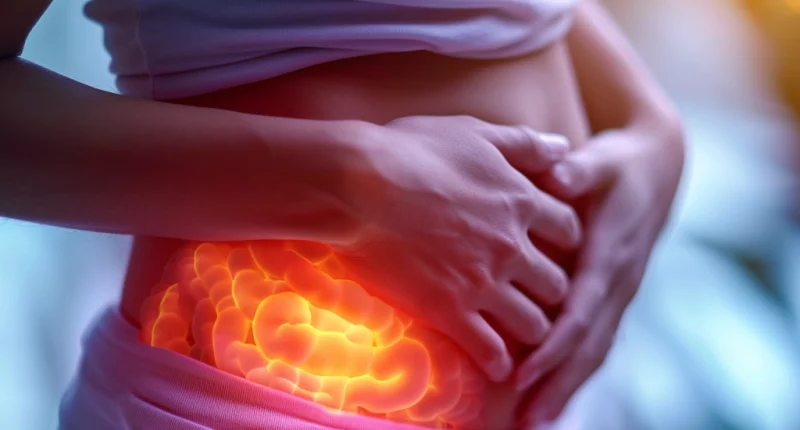
Cholecystitis, also known as gallbladder inflammation, is a condition that affects the gallbladder, a small pear-shaped organ located beneath the liver on the right side of the abdomen. This organ plays a crucial role in storing and distributing bile, a digestive fluid released into the small intestine.
The primary cause of cholecystitis is often the presence of gallstones obstructing the tube leading out of the gallbladder. These gallstones can lead to a backup of bile, resulting in inflammation. However, other factors can trigger cholecystitis, including issues with the bile ducts, the presence of tumors, serious illnesses, and specific infections.
When cholecystitis is left untreated, it can progress to severe and potentially life-threatening complications, such as a ruptured gallbladder. In many cases, the recommended treatment for cholecystitis involves surgical removal of the gallbladder to alleviate the symptoms and prevent further complications.
Cholecystitis Classification
- Acute, characterized by a sudden and urgent onset.
- Chronic, marked by a slow and long-lasting progression.
- Calculous, linked to the presence of gallstones.
- Acalculous, unrelated to the presence of gallstones.
Causes of cholecystitis
The most common cause of chronic or acute cholecystitis is the presence of gallstones that obstruct your bile ducts. Gallstones, which are solidified remnants of bile, typically form within the gallbladder but can occasionally travel and become lodged in a bile duct or the gallbladder’s opening. This obstruction results in the backup of bile into the gallbladder, creating an environment conducive to infections.
Acute cholecystitis is often triggered by a gallstone that completely obstructs the bile flow from the gallbladder. As the gallbladder swells, the condition progressively worsens. On the other hand, chronic cholecystitis can be caused by a drifting gallstone that only partially obstructs the gallbladder, leading to intermittent symptoms, often occurring after consuming a heavy meal.
However, gallstones aren’t the only culprits of gallbladder inflammation. Other factors can lead to the slowing and backup of bile into the gallbladder, such as:
- Biliary Stricture: Chronic inflammation in the bile ducts can eventually result in scar tissue formation, narrowing the bile ducts and obstructing bile flow.
- Biliary Dyskinesia: This functional gallbladder disorder affects gallbladder motility, causing insufficient contractions that prevent the proper expulsion of bile.
- Tumor: Although rare, tumors in the gallbladder or bile ducts can obstruct bile flow, leading to cholecystitis.
- Bile Stasis: Chronic liver disease or long-term parenteral nutrition can stall bile flow, especially when nutrition is administered intravenously, bypassing the digestive system’s need for ile.
These conditions tend to develop gradually, making them more likely to cause chronic cholecystitis. However, they can progress over time, and chronic acalculous cholecystitis may evolve into acute acalculous cholecystitis.
Additional potential causes of gallbladder inflammation include:
- Ischemia: Reduced blood flow to the gallbladder can result in either chronic or acute inflammation, depending on its severity. Ischemia is often a response to critical illness or trauma and can also occur due to vascular occlusion or inflammation of the blood vessels (vasculitis).
- Severe Illness: Extremely severe illnesses can cause damage to blood vessels and decrease blood flow to the gallbladder, ultimately leading to cholecystitis.
- Infection: While less common, bacterial infections can affect the gallbladder or bile ducts even without an obstruction trapping bacteria. Individuals with weakened immune systems are more susceptible to these infections, which can irritate and erode gallbladder tissue.
Symptoms of cholecystitis
Common symptoms of cholecystitis include:
- Severe upper abdominal pain that can radiate to the right shoulder or back, often mistaken for chest pain or a heart attack. This pain intensifies after a large or fatty meal and may be sharp, dull, or crampy.
- Nausea, vomiting, and abdominal tenderness are also typical.
- Fever occasionally indicates infection or severe inflammation, though less common in older individuals.
Chronic cholecystitis symptoms are milder and intermittent, typically occurring after heavy meals, leading to abdominal discomfort and bloating.
How to prevent cholecystitis
To prevent cholecystitis and reduce the risk of gallstones, consider these measures:
- Gradual Weight Loss: Avoid rapid weight loss, which can increase gallstones’ likelihood.
- Maintain Healthy Weight: Stay within a healthy weight range by adjusting your diet to lower calorie intake and increasing physical activity.
- Opt for a Balanced Diet: Reduce the risk of gallstones by choosing a diet rich in fruits, vegetables, whole grains, and fiber and low in saturated fats.
While there is some evidence supporting the preventative effects of a healthy lifestyle, diet, regular physical activity, and maintaining an ideal body weight, it should be noted that the strength of this evidence is limited.
Cholecystitis diagnosis
Diagnosing cholecystitis involves a thorough evaluation conducted by your healthcare provider. The diagnostic process typically begins with discussing your symptoms and medical history, followed by a physical examination. To confirm the presence of cholecystitis, various tests and procedures are employed:
- Blood Tests: Your healthcare provider may request blood tests to identify signs of infection.
- Imaging Techniques: Several imaging methods are available to visualize the gallbladder and bile ducts, including abdominal ultrasound, endoscopic ultrasound, CT scan, and MRCP (magnetic resonance cholangiopancreatography). These imaging studies assist in detecting cholecystitis indicators and the presence of gallstones.
- Bile Flow Evaluation: A hepatobiliary iminodiacetic acid (HIDA) scan is utilized to monitor the production and flow of bile from the liver to the small intestine. This scan involves injecting a radioactive dye that binds to bile-producing cells, allowing for the identification of any obstructions in the bile ducts.
In addition to these assessments, your healthcare provider may conduct specific tests such as a complete blood count (CBC), liver function tests, and an evaluation of tenderness in your abdomen (Murphy’s sign) to facilitate the diagnosis of cholecystitis.
Treatment for cholecystitis
The management of cholecystitis involves a comprehensive approach, typically administered within a hospital setting. This treatment protocol comprises:
Surgery
The primary and most prevalent treatment for cholecystitis is the surgical removal of the gallbladder, known as cholecystectomy. Gallstones and various gallbladder-related issues often underlie cholecystitis.
Surgery may remain necessary even after the resolution of acute symptoms, as both chronic and acute cholecystitis have a propensity to recur. Repeated episodes of gallbladder inflammation can result in cumulative damage.
Cholecystectomy is performed in more than 612 hospitals worldwide (https://doctor.global/results/diseases/cholecystitis). The cost fluctuates depending on the location. For instance, in Poland, the cost of gallbladder removal ranges from 2000 to 6000 EUR. (https://doctor.global/results/europe/poland/all-cities/all-specializations/diseases/cholecystitis).
Supportive Care
- Administering intravenous (IV) fluids to ensure adequate hydration and provide rest for the digestive system.
- Prescribing IV antibiotics to address or prevent potential infections associated with cholecystitis.
- Providing IV pain relief to alleviate the common discomfort experienced by individuals with this condition.
Alternative Procedures
In cases where immediate surgery is not feasible or recommended, alternative approaches may be considered:
- Gallbladder drainage (cholecystostomy) may be performed to eliminate infection when surgery is not viable. This may involve drainage through the abdominal skin (percutaneous) or an endoscopic procedure.
- Endoscopic retrograde cholangiopancreatography (ERCP) is a procedure that employs dye to highlight the bile ducts. During ERCP, instruments can be employed to remove obstructions caused by stones in the bile or cystic ducts.
Following gallbladder removal surgery (cholecystectomy), your body will adapt to the absence of the gallbladder over several weeks to months. During this transitional phase, you may experience temporary symptoms such as reduced bile flow, resulting in sensations of pressure or discomfort in the biliary system. Some individuals may also encounter difficulty digesting fats, potentially leading to diarrhea.
In summary, the treatment for cholecystitis aims to alleviate symptoms, resolve inflammation, and prevent future occurrences of gallbladder-related issues.


