Diagnostic & Treatment Management of Colorectal Cancer Disease
Algorithm of diagnosis
What evaluations do CRC patients undergo to identify the best treatment strategy?
It is crucial to understand – both for a patient and a physician – the stage and clinical presentation of the disease so patient treatment and support are of total value. Additionally, in women, the diagnostic algorithm helps to rule out the presence of synchronous breast, ovarian, and endometrial cancers.
The diagnostic interventions include:
- Clinical examination: includes palpation (touching) of the abdomen and rectal examination. By doing so, the doctor determines whether the tumor has caused the liver to enlarge and whether it has caused excess fluid in the abdomen, called ascites. The rectal examination serves to identify if there are any signs of blood in the area.
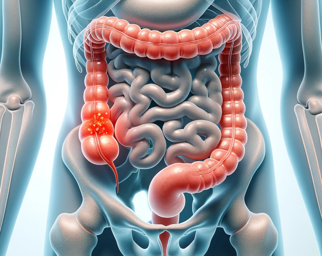
- Endoscopy: allows the doctor to inspect the interior of the bowel for abnormal formation or growths in the inner lining of the intestine using a fine-lighted tube with camera and biopsy tools. If any abnormal lesion is found, the doctor can take a piece of it for histopathological (microscopic) examination. Endoscopy can be performed in different areas, by inserting a short rectoscope, longer sigmoidoscope, or a flexible colonoscope to investigate a target part of the large intestine precisely and carefully. Tumors found within 15 cm of the anus are classified as rectal cancer, whereas any further away from the anus location are called colon tumors. When a rectal tumor is found during a rectoscopy, a complete colonoscopy is also required to check if the pathology resides locally or distributed through the gut.
- Radiological investigation: if the endoscopy is challenging to perform, for example, in obstructive tumors, a virtual colonoscopy may be an option. This examination involves a computer tomography (CT) scan of the abdomen, after which a computer produces 3-D images from the interior wall of the large intestine.
An outdated but occasionally used radiological option is a double contrast barium enema with barium sulfate (a chalky liquid commonly used in radiological examinations), and air are introduced into the colon via the anus, typically when the right-sided part of the colon is under investigation.
For the endoscopy and virtual colonoscopy, adequate bowel preparation is needed.
Additional radiological techniques may be used to determine the level of the tumor spread and the presence of metastases. They include a CT scan of the chest, ultrasound of the liver, and endoscopic ultrasound. While nuclear magnetic resonance imaging (MRI) of the rectum is a routine diagnostic procedure in rectal cancer, it stays optional in colon cancer. Positron emission tomography (PET) is another way to visualize metastasis in high resolution, but PET scans are not performed routinely due to their cost.
- Laboratory tests: routine blood sample measurements give information on complete blood count and liver/kidney function. Tumor markers monitoring helps to identify a level of a specific protein called carcinoembryonic antigen (CEA), which is sometimes elevated in this pathology. If CRC patients have high CEA levels at initial diagnosis, observing this marker level during and after treatment is wise.
- Histopathological examination: means laboratory investigation of the tumor tissue under a microscope on the biopsy or polyp obtained via endoscopy. As a result, the doctor will have information on specific mutations (such as RAS, BRAF, MLH1, CIN, MSI, etc.) within tumor cells sensitive to target therapy (molecular profiling). Also, the doctor performs histology-assessed tumor staging from 0 to IV, which helps to understand the disease’s prognosis and possible regional or metastatic risk.
Treatment routes
What is an appropriate treatment for different CRC stages?
Surgery is the most effective one-step maneuver to treat CRC forms that have not spread to distant sites. Radiation therapy and/or chemotherapy may also be options for people who aren’t healthy enough for surgery or for when complete resection is not possible due to tumor location.
Treating stage 0 CRC
The stage 0 colon tumors have not grown beyond the inner lining of the colon. That is why surgery to take out the cancer via excision of the polyp (polypectomy) through the intestinal lumen (colonoscopy approach) is often the only treatment needed. This procedure is called endoscopic mucosal resection (EMR) or transanal minimally invasive surgery (TAMIS).
EMR is performed in more than 690 clinics worldwide with an approximate cost of:
- Spain – $2.5 K
- Israel – $7.3 K
- France – $3.9 K
- Turkey – $1.8 K
- Saudi Arabia – $1.4 K
Treating stage I CRC
A more advanced surgical approach can be utilized when the cancer has grown deeper through the intestinal wall but has not spread outside the colon wall itself or into nearby lymph nodes. In the case of polyp-formed CRC stage I, the polypectomy (polyp removal) during colonoscopy via EMR or local excision with a small amount of surrounding healthy tissue on the colon wall via TAMIS is performed. If no cancer cells are at the edges (margins) of the removed piece, no other treatment may be needed.
However, there are following situations when surgical removing a small part of the colon (partial colectomy ≥ 5 cm on either side of the tumor) is a standard treatment in advanced CRC stage I:
- if the tumor presents as high-grade (highly abnormal cells on histology),
- if there are cancer cells at the edges of the polyp, or if the polyp couldn’t be removed completely,
- if the polyp had to be removed in many pieces.
If only part of the colon is removed, it’s called a hemicolectomy, partial colectomy, or segmental resection. The surgeon takes out the part of the colon with the cancer and a small segment of the normal colon on either side. Usually, about one-fourth to one-third of the colon is removed, depending on the size and location of the cancer. A colectomy can be done in 2 ways:
- open colectomy when the surgery is done through a single long incision (cut) in the abdomen (belly);
- laparoscopic-assisted colectomy, also known as minimally invasive surgery – through 3-4 smaller incisions (0,5-2 cm length) in the belly with the use of special manipulation tools and a small camera. Due to minor body damage, patient recovery in this option is much faster, while overall survival rates and the chance of cancer returning are the same compared to open colectomy.
After the operation, the colon’s remaining sections are reattached in three ways: end-to-end, side-to-side (prevents lumen narrowing), or end-to-side (connects smaller lumen with the larger one). In the advanced CRC stage, end-to-end and side-to-side connections (anastomoses) are options of choice. At least 12 nearby lymph nodes are also removed so they can be checked for cancer.
Treating stage II CRC
The CRC at stage II penetrates the intestinal wall and may invade nearby tissue. Patients in this group receive surgery as a standard cure method – a partial colectomy along with nearby lymph nodes removal. Another name for this operation is En bloc colonic and mesentery resection since cancer lesions in the intestinal wall and infiltrated fat tissue are resected without breaking the tumor capsule.
Suppose the involved area is too large to perform an anastomosis on the resting bowel ends immediately after the resection. In that case, the surgeon will move one end of the intestine through an opening in the abdominal wall and connect it to a bag or pouch. This is called colostomy and it’s done so that stools that would normally move through the intestine to the rectum instead pass through the opening in the abdomen into the pouch. The colostomy bag must be manually emptied. The colostomy is often only used as a short-term solution and consequently is replaced by ileocolonic anastomosis, which re-connects the small intestine and colon.
If a CRC stage II tumor looks very abnormal when viewed closely in the lab, or it has grown into nearby blood or lymph nodes, or there is a blocked (obstructed) lumen / perforated (hole) colon wall, the neoadjuvant chemotherapy for 3-6 months is recommended, including capecitabine or fluoropyrimidine (5-FU) treatment.
The medication treatment starts as adjuvant therapy within eight weeks after the surgery at stages II-IV, when the tumor can involve the whole depth of the bowel, raising intestine wall perforation awareness.
Treating stage III CRC
Stage III colon cancer is widespread to nearby lymph nodes, which is why surgeons often perform partial colectomy, followed by 3-6 months adjuvant treatment (5-FU, oxaliplatin, capecitabine) to prevent further spreading of the disease without mutations dMMR or MSI-H. If these mutations appear, neoadjuvant immunotherapy (pembrolizumab, etc.) is recommended.
Radiation therapy may accompany the surgery when some advanced stage III tumors are found to be attached to a nearby organ:
- before the surgery, radiation therapy helps to shrink a tumor and make it easier to remove;
- after the surgery – double-check for cancer cell killing in a region;
- during the surgery – intraoperative radiation therapy (IORT) – to clean the area from possible tumor cells left.
Treating stage IV CRC
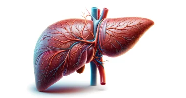
The challenge with terminal stage IV CRC is that it is a metastatic tumor (mCRC) that involves the liver, lungs, and bones. That’s why the surgery bears only a palliative goal, meaning the partial colectomy or the total colectomy (removing all the colon) would not have a decisive effect on the patient’s recovery.
Total colectomy can be done in more than 690 clinics worldwide for an approximate price of:
- Switzerland – $21.3 K
- United States – $50 K
- Thailand – $11.9 K
- Turkey – $7.7 K
- UAE – $13.2 K
The number of operable cases in CRC decreases with stage growth. Starting from stage I CRC– about 90% of patients are surgery-eligible – to metastatic CRC (mCRC) stage IV, where due to comorbidities, there is about a 20% chance to be eligible for radical surgery.
At CRC stage IV, radiation therapy, chemotherapy, and immunotherapy are the first-line treatments.
Radiation therapy includes three types:
- External-beam radiation therapy (EBRT) – helps to focus the radiation beam on the cancer from a machine outside the body. There are techniques, such as three-dimensional conformal radiation therapy (3D-CRT), intensity modulated radiation therapy (IMRT), and stereotactic body radiation therapy (SBRT), have been shown to treat lungs and liver CRC metastases;
- Internal radiation therapy (brachytherapy) – treats rectal cancer since the radiation source is put inside the rectum next to or into the tumor;
- Radioembolization – means injecting tiny beads (called microspheres) coated with radioactive yttrium-90 (Y-90) into the hepatic artery. The beads lodge in the blood vessels near the metastasis in the liver, where they give off small amounts of radiation to the tumor site for several days.
Chemotherapy treatment of peritoneum (inner wall of the abdomen) metastatic lesions in mCRC includes:
- Hyperthermic Intraperitoneal Chemotherapy (HIPEC) uses high-concentration drug mitomycin-C combined with hyperthermia 39-43°C and high-pressure flow;
- Pressurized Intraperitoneal Aerosolized Chemotherapy (PIPAC) uses high pressure to push chemo drug (oxaliplatin) into metastatic cells.
Mutation-specific approach for CRC (immunotherapy)
Are there any personalized medicine options?
A few options for standard treatment-resistant mutation-based personalized approach as a first-line therapy were developed in the last decade. The immunotherapy drugs (bevacizumab, cetuximab, panitumumab, etc.) target specific molecular proteins from a group called Growth Factors, which provide cancer rapid growth and spread when genetic mutations trigger its overproduction. The medications block the ability of cells to produce the growth factors, thus shrinking the tumor.
Follow-up & surveillance strategy
How do you make sure the cancer will not return?
Monitoring of CRC blood markers such as carcinoembryonic antigen (CEA) may help to catch a cancer recurrence after initial treatment at an early stage.
Localized Colon Cancer stage 0-III surveillance program:
- when surgery is not possible due to significant comorbidities, surveillance colonoscopy within six months after polyp removal with close oncological follow-up, including a CT scan to detect lymph node recurrences, is recommended;
- if the surgery went successfully, repeat colonoscopy in 1-2 years and 3-5 years thereafter;
- CEA determination and physical examination every 3-6 months for three years and every 6-12 months at years 4 and 5 after surgery;
- CT scans of the chest and abdomen every 6-12 months for the first three years for high-risk recurrence stage II-III CRC patients.
Metastatic Colon Cancer stage III-IV surveillance program:
- for patients receiving active treatment, a CT scan or MRI plus CEA measurement every 8-12 weeks is recommended;
- after radically resected metastatic disease – CT or MRI plus CEA levels every three months during the first two years and every six months thereafter.
Have a look at the related article “Colorectal Cancer – Overview“.
Hereunder you can also find the short video explanation of colorectal cancer disease overview and treatment:
Colorectal Cancer Treatment FAQ
- Clinical Examination: Palpation of the abdomen and rectal examination to check for tumor-related changes like liver enlargement or ascites.
- Endoscopy: Visual inspection of the bowel's interior using a fine-lighted tube with a camera and biopsy tools to identify abnormal formations and obtain tissue samples.
- Radiological Investigations: Techniques like CT scans, virtual colonoscopy, and MRI to visualize the tumor and check for metastases.
- Laboratory Tests: Blood tests to measure liver/kidney function and tumor markers like carcinoembryonic antigen (CEA).
- Histopathological Examination: Microscopic analysis of biopsy samples to identify specific mutations and stage the tumor.
- Stage 0: Surgery to remove the tumor via endoscopic mucosal resection (EMR) or transanal minimally invasive surgery (TAMIS).
- Stage I: Surgery to remove the tumor and possibly a small portion of the colon (partial colectomy). Additional treatments depend on the tumor's characteristics.
- Stage II: Surgery (partial colectomy) along with nearby lymph nodes removal. Chemotherapy may be recommended if the tumor shows high-risk features.
- Stage III: Surgery followed by chemotherapy (5-FU, oxaliplatin, capecitabine) and possibly radiation therapy.
- Stage IV: Palliative surgery, radiation therapy, chemotherapy, and immunotherapy to manage metastatic disease.
- Localized Colon Cancer (Stage 0-III): Surveillance colonoscopy within six months post-treatment, then at 1-2 years and every 3-5 years thereafter. Regular CEA measurements and physical exams every 3-6 months for the first three years, then every 6-12 months for years 4 and 5. CT scans every 6-12 months for high-risk patients.
- Metastatic Colon Cancer (Stage III-IV): For active treatment patients, CT or MRI scans plus CEA measurements every 8-12 weeks. After radically resected metastatic disease, scans and CEA levels are checked every three months for the first two years and every six months thereafter.
- Stage 0: Surgery involves removing the cancerous polyp via EMR or TAMIS.
- Stage I: Surgery may involve a simple polypectomy or partial colectomy if the tumor is more advanced.
- Stage II: Surgery typically involves a partial colectomy with lymph node removal.
- Stage III: Surgery is combined with chemotherapy and possibly radiation therapy.
- Stage IV: Surgery is usually palliative, aiming to relieve symptoms rather than cure the disease.