Femoral fracture
Definition
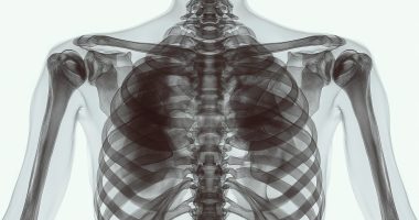
Femur fractures account for about 6% of all bone fractures. Three main groups of femur fractures are distinguished: fractures of the upper end of the femur, diaphyseal fractures, and fractures of the lower end. Depending on the location of the hip fracture, it may manifest itself in pain, limitation of hip mobility, shortening, and deformation of the injured limb. In an open fracture, significant blood loss is possible. The primary way of diagnosing hip fractures is radiography. In intra-articular hip fractures, an MRI of the joint is additionally performed. Hip fracture treatment consists of positioning the fragments and their fixation with spokes, three-blade nail nails, and external fixation apparatus; skeletal traction is used if indicated.
General information
Hip fractures account for about 6% of all bone fractures. There are three main groups of hip fractures:
- Fractures of the upper (proximal) end of the femur. This group includes femoral neck fractures and acetabular fractures;
- Diaphyseal fractures of the femur (fractures of the body of the femur);
- fractures of the lower (distal) end of the femur.
The listed groups of hip fractures differ in the mechanism of injury, clinical symptomatology, treatment tactics, and long-term prognosis.
Anatomy
Like all other tubular bones, the femur consists of a body (diaphysis) and two ends (epiphyses). The head is located in the upper part of the femur, which enters the articular socket of the pelvic bones to form the hip joint.
Below the head of the femur is the thinner neck. The neck of the femur joins the body at an angle. There are protrusions (the greater and lesser trochanters) on the outside of the femoral neck. The lower end of the femur expands to form two condyles (inner and outer). With their articular surfaces, the condyles border the tibia and patella to form the knee joint.
Causes of hip fractures
Hip fractures usually result from direct trauma (fall, impact) or indirect trauma (twisting, bending). The cause of damage can be a fall from a height, a car accident, or industrial or sports trauma. Young and middle-aged people are more often affected.
Direct trauma results in transverse, oblique, and splinter fractures of the femur, while indirect trauma results in screw fractures. Many muscles attached to the femur affect the fragments in a hip fracture. The muscles pull the fragments apart, causing them to dislocate. The direction of displacement depends on the level of the fracture.
Distal fractures
Femoral condyle fractures occur due to a fall or a direct blow to the knee joint. They may be accompanied by the dislocation of the fragments. Older adults are more often affected.
One or both condyles may be fractured. The fragments in a femoral condyle fracture are characteristically displaced upward and sideways. The fracture line runs inside the joint. Blood from the fracture site pours into the joint, causing hemarthrosis.
Symptoms
A patient with a hip fracture complains of severe pain at the site of injury. Swelling, hemorrhage, deformity of the limb, and abnormal mobility are observed in the area of the fracture. The leg is usually shortened. A fracture of the femur may be accompanied by damage to a nerve or a large vessel. Due to sharp pain and marked blood loss, traumatic shock is possible.
In distal femur fractures, the patient complains of sharp pain in the knee and lower thigh. Movement in the joint is limited and sharply painful. The knee joint is enlarged in volume. A fracture of the external condyle is accompanied by tibia deviation to the outside. With a fracture of the internal condyle, the patient’s tibia is deviated to the inside. MRI of the knee joint, along with radiography, is also used to diagnose femoral condyle fractures.
Treatment
As first aid, the injured limb should be fixed by applying a splint. The patient is given an anesthetic, covered with ablanket, and transported to a hospital.
There is a risk of traumatic shock in hip fractures. Preventive antishock measures include adequate anesthesia and blockade of the fracture site. In case of significant blood loss, blood transfusion, and blood substitutes are administered. A plaster bandage at the initial stage of treatment is not used since, with its help, it is impossible to keep the fragments in the correct position. Skeletal traction, external fixation devices, and surgery (osteosynthesis) are used as the main methods of treatment.
Contraindications to surgical treatment for hip fracture are severe concomitant diseases, infected wounds, and the general serious condition of the patient as a result of combined trauma. If there are contraindications to surgery, skeletal traction is indicated for 6-12 weeks. The spica for skeletal traction is inserted through the femoral condyles or tibial tuberosity. The patient is placed on a shield; the injured leg is placed on a Belair splint. The amount of weight in hip fracture is determined by the level of the fracture and the nature of displacement.
The load can be increased in young patients with well-developed muscles. The average weight at the beginning of treatment is about 10 kg. As the displacement is eliminated, the weight is reduced. After the traction is removed, a plaster cast is applied to the injured limb for up to 4 months. With conservative treatment, the knee and hip joints remain immobile for a long time. Surgical treatment increases the patient’s mobility and prevents the development of contractures. Surgery is performed after normalization of the patient’s condition. Osteosynthesis is performed using plates, pins, and rods.
On admission, a local anesthetic (novocaine) is injected into the fracture area for pain relief. Further treatment tactics are determined by the traumatologist according to the level of the fracture and the patient’s general condition. In intra-articular fractures, surgical treatment is preferable, providing fusion in 70% of cases. Contraindications to surgery are severe comorbidities and advanced age of the patient.
The advanced age of patients with femoral neck fractures and the presence of concomitant diseases cause a high incidence of complications during prolonged bed rest. Patients often develop bedsores and pneumonia. Thromboembolism is possible. Due to the large number of complications, the choice of treatment tactics for such patients should adhere to the general principle – to ensure maximum mobility of the patient in combination with possible immobilization of the limb in these conditions. If the patient’s condition allows surgery, fixation with a three-bladed nail or bone autoplasty is performed.
Subsequently, patients with femoral neck fractures may develop a false joint or aseptic necrosis of the femoral head, for which hip replacement is indicated. Skeletal traction is used for hip fractures for eight weeks. After the traction is removed, a plaster cast is applied. Stepping on the injured leg is allowed after 3-4 months. Surgery for vertebral fractures can shorten the treatment period and increase the patient’s mobility. Osteosynthesis is performed with a three-lobed nail, plates, or screws. The total load on the leg is allowed after 6-10 weeks.
All these treatment options are available in more than 900 hospitals worldwide (https://doctor.global/results/diseases/femoral-fracture). For example, Intramedullary nailing can be performed in following countries for an approximate price of:
Turkey$2.8 K in 29 clinics
China$10.5 K in 8 clinics
Germany$10.7 K in 43 clinics
Israel$13.4 K in 14 clinics
United States$15.9 K in 15 clinics
Prognosis
The femoral neck is not covered with periosteum. The blood supply to the neck and head is impeded, so femoral neck fractures do not heal well. Due to insufficient nutrition, complete fusion only occurs in some cases. Over time, the fragments are partially fixed by a dense connective tissue scar. So-called fibrous fusion occurs. The prognosis for femoral neck fractures is worse the higher the fracture line is located. Without surgical treatment, the outcome of “high” fractures of the femoral neck is often disability.
The acetabular region is well supplied with blood, creating favorable conditions for forming a full-fledged bone callus. In most cases, if adequately treated, acetabular fractures of the femur heal well without surgery.