Flat foot
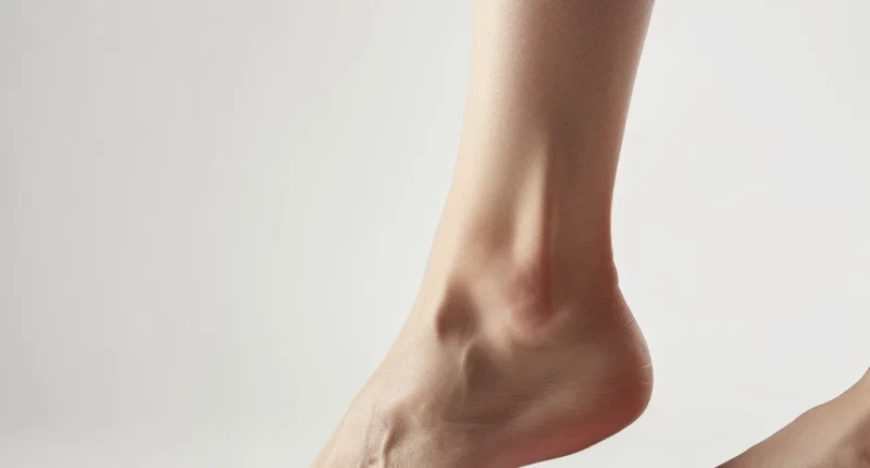
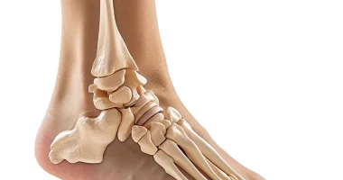
Flatfoot is a flattening of the longitudinal and transverse arch of the foot. This condition results in incorrect function of the musculoskeletal compartment and the mobile joints of the foot.
Overview
Flatfoot, commonly known as flat feet, is a condition prevalent in about half of preschoolers, with its frequency slightly decreasing to 15% in schoolchildren and averaging 15-20% in adults. This condition is characterized by one or both feet having minimal or no arch, causing the pads of the feet to press fully into the ground when standing. While some children exhibit visible arches when lifting their feet, the absence of arches is typical in this age group.
Risk factors for developing flat feet include age-related characteristics, as younger children are more likely to be diagnosed with this condition. Other factors include constitutional features, weak connective tissues such as ligaments, genetic predisposition, and early onset of wearing shoes. Children should take their first steps barefoot or in socks for proper foot proprioceptive innervation.
In the context of foot development, flatfoot is often considered normal up to the age of 5-7 years, assuming no accompanying symptoms or adverse effects. However, children with this condition should be under dynamic observation by an orthopedic traumatologist due to increased risks of pain syndrome, lameness, sensory and trophic disorders, and degenerative-dystrophic joint issues.
For most people, flat feet do not cause significant problems. However, if they lead to pain or other issues, there are effective treatment options available.
Flat Foot classification
- Flexible Flat Feet: This is the most prevalent type, where arches are visible when not standing but disappear underweight. It typically emerges in childhood or adolescence, affecting both feet, and may worsen over time due to stretching or damage to the tendons and ligaments in the arches.
- Rigid Flat Feet: In this type, no arches are present, whether standing or sitting. It often starts in the teens and can progress with age, leading to pain and difficulty moving the feet up, down, or side-to-side. It can affect one or both feet.
- Adult-Acquired (Fallen Arch): This occurs when the arch collapses suddenly in adulthood, often due to inflammation or a tear in the posterior tibial tendon. It leads to an outward turning of the foot and can be painful, typically affecting just one foot.
- Vertical Talus: A congenital disability in some babies where the talus bone is mispositioned, preventing arch formation. The foot’s bottom looks like the bottom of a rocking chair.
Depending on the height of which arch of the foot is reduced, flatfoot is divided into the following forms:
- Longitudinal – the longitudinal arch is flattened;
- Transverse – the transverse arch is flattened;
- Combined – a combination of the above two types;
Causes of Flatfoot
Flatfoot can be both a genetic condition and a result of various environmental factors. While some individuals inherit this condition, for others, arches don’t develop significantly during childhood, leading to flat feet. Additionally, certain life stages and health conditions can increase the risk of developing flat feet. These include:
Genetic and Developmental Factors:
- Inherited traits in families.
- Congenital anomalies like a vertical talus or “rocking foot,” where the talus bone is abnormally positioned.
- Tarsal coalitions, a prenatal condition where foot bones abnormally fuse together.
Medical Conditions:
- Achilles tendon injuries.
- Cerebral palsy.
- Obesity.
- Rheumatoid arthritis
- Diabetes.
- Down syndrome.
- High blood pressure.
- .Pregnancy.
Physical Stress and Lifestyle:
- Trauma or broken bones in the feet.
- Increased body weight exceeding normal limits.
- A sedentary lifestyle with minimal physical activity.
External Influences:
- Wearing non-anatomical or poorly supportive footwear.
- Adverse outcomes from medical interventions on the foot.
These diverse causes contribute to the formation of flat feet, which can either be a flexible type commonly seen in children or a rigid form often resulting from congenital or acquired foot abnormalities.
Flatfoot diagnosis
Clinical criteria are used to assess the severity of foot deformity and several instrumental methods of examination (plantography, radiography, radiometry). In complex clinical cases, multislice computed tomography or nuclear magnetic resonance examination may be indicated to establish the diagnosis.
- The basic diagnostic method is the examination of the patient, which allows you to examine the state of the skeleton and identify possible violations of the functional state. When conducting the examination, the doctor considers the age category (child, adult male/female) and the nature of deformation under load and in the absence of it. Studies confirm that the development of the vault of the foot in the longitudinal direction continues during the first ten years of life, and the frequency of the development of flat feet has an inverse relationship with the child’s age and a direct relationship with body weight. It is recommended to resort to clinical and manual tests to determine the mobility of the deformity.
- One of the most accessible instrumental methods for determining the degree of flattening of the longitudinal arch of the foot is plantography. Unlike radiography, plantography does not carry a radiation burden to the patient. Many researchers evaluate the results using the Staheli and Chippaux Smirak index (CSI).
- Imaging Tests: To assess the bones and tissues of the foot more closely and to rule out other conditions, imaging tests such as X-rays, MRI (Magnetic Resonance Imaging), or CT (Computed Tomography) scans may be used. These tests provide detailed images of the foot structure, helping in the identification of flatfoot and its severity.
Flatfoot treatment
The treatment of flat feet usually involves conservative methods, though surgery might be necessary in certain cases.
Conservative Treatment Methods
Orthotic Devices: These are commonly employed in treating flat feet and include:
- Special inserts to correct the longitudinal and transverse arches of the foot.
- Heel inlays for additional support.
- Toe separators for improved alignment.
Custom orthotics are particularly effective in enhancing foot functionality, alleviating pain, and thus improving the patient’s overall quality of life. They also help increase physical activity endurance and redistribute the load from overstrained muscles to more relaxed ones.
Physical Therapy: An integral part of the conservative treatment plan, physical therapy exercises significantly benefit the muscle compartment of the foot. Types of therapeutic exercises include:
- Walking routines.
- Passive stretching exercises.
- Manual therapy techniques.
- Mechanical therapy methods.
Such exercises are designed to strengthen the foot muscles, enhance muscle tone distribution across different foot areas, and positively impact the overall condition of the lower extremities.
Surgical Intervention
Surgery for flat feet is recommended in the following cases:
- lack of therapeutic results from conservative management of the patient;
- long-term persistent pain syndrome.
Modern orthopedics has various methods of surgical treatment. They aim to restore the correct anatomy of the foot and improve its functional state.
On Doctor.Global there are 768 clinics worldwide where flat foot can be treated (https://doctor.global/results/diseases/flat-foot). For example, the cost of correcting foot deformities in Poland ranges between 1352 USD and 3463 USD (https://doctor.global/results/europe/poland/all-cities/all-specializations/diseases/flat-foot).
Flatfoot prevention
Engaging in adequate physical activity during childhood and adolescence is crucial to prevent the development of flat feet. Beneficial activities include exercises using a stick, ball, jump rope, and hula-hoop.
Key preventative exercises include:
- Rolling a jump rope with the foot, moving it from the back to the front and vice versa.
- Flexing and extending the toes as if mimicking a caterpillar’s movement.
- Walking on heels.
- Walking on the sides of the feet while keeping the body balanced.
- Toe raises, ensuring feet are kept shoulder-width apart.
Along with these exercises, incorporating orthopedic insoles, self-massage, swimming, and aqua aerobics into the routine is recommended. These activities positively influence the musculoskeletal structure of the foot, aiding in the prevention of flat feet.


