Degenerative disc disease (DDD)
Definition
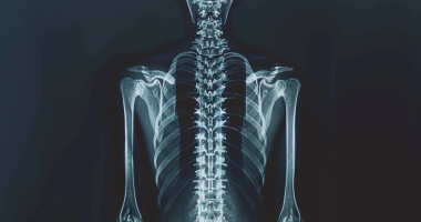
Degenerative disc disease (DDD), also called osteochondrosis of the spine, is a chronic disease in which degenerative changes occur in the vertebrae and the intervertebral discs between them. Depending on the location of the spine affected, cervical osteochondrosis, thoracic osteochondrosis, and lumbar osteochondrosis are differentiated. Diagnosing osteochondrosis of the spine requires radiography and, in the case of its complications (for example, herniated disc) – an MRI of the spine. Medication methods are widely used in treating osteochondrosis of the spine: reflexology, massage, manual therapy, and physiotherapy.
Etiology and pathogenesis
Osteochondrosis of the spine develops to some degree in all people as they age and is one of the body’s aging processes. Earlier or later, atrophic changes occur in the intervertebral disc, but trauma, disease, and various overloads of the spine contribute to the earlier occurrence of osteochondrosis. Osteochondrosis of the cervical spine and osteochondrosis of the lumbar spine are the most common.
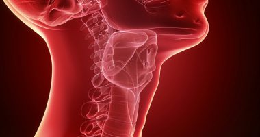
It is believed that the main point in the occurrence of osteochondrosis is the constant overloading of the spinal-motor segment, which consists of two neighboring vertebrae with an intervertebral disc located between them. This overloading can result from motor stereotypes – posture and individualized manner of sitting and walking. Violations of posture, sitting in the wrong position, and walking with an uneven spinal column cause additional stress on the spine’s discs, ligaments, and muscles. The process can be aggravated due to the peculiarities of the spine’s structure and insufficient tropism of its tissues due to hereditary factors. Most often, malformations in the structure are found in the cervical spine (Kimmerle anomaly, craniovertebral anomalies, Chiari anomaly) and lead to vascular disorders and early signs of osteochondrosis of the cervical spine.
Lumbar osteochondrosis is more often associated with overloading the lumbar disc by bending over and lifting weights. A healthy intervertebral disc can withstand significant loads due to the hydrophilicity of the pulp nucleus located in its center.
In osteochondrosis, the pulp nucleus loses its hydrophilic properties. That occurs due to disorders in its metabolism or an insufficient supply of necessary substances. As a result, the intervertebral disc becomes flat and less elastic, and radial cracks appear in its fibrous ring under load. The distance between neighboring vertebrae decreases, and they shift in relation to each other, with displacement also occurring in the facet (arch) joints that connect the vertebrae.
Due to the displacement of vertebral bodies, the capsules of the facet joints are stretched, and the altered intervertebral disc no longer fixes the bodies of neighboring vertebrae so firmly. Vertebral segment instability is formed. Due to instability, it is possible to pinch the root of the spinal nerve with the development of radicular syndrome. Osteochondrosis of the cervical spine often occurs during head turns, while osteochondrosis of the lumbar spine – occurs during tilts of the torso.
A disc herniation occurs when the disc is displaced backward, the posterior longitudinal ligament is torn, and part of the disc protrudes into the spinal canal. If the pulposus nucleus of the disk is pressed into the spinal canal, then such a hernia is called ruptured. The severity and duration of pain in such a herniation are much greater than in a non-torn herniation. A herniated disc can cause radicular syndrome or spinal cord compression.
Osteochondrosis causes bone overgrowth, forming osteophytes, which are bony growths on the vertebral bodies and spines. Osteophytes can also compress the spinal cord (compression myelopathy) or cause radicular syndrome.
Symptoms of osteochondrosis of the spine
The main symptom of osteochondrosis of the spine is pain. Pain can be sharp with high intensity; it increases at the slightest movement in the affected segment, forcing the patient to take a forced position. Thus, with osteochondrosis of the cervical spine, the patient holds his head in the least painful position and cannot turn it; with osteochondrosis of the thoracic spine, the pain increases even with deep breathing, and with osteochondrosis of the lumbar spine, the patient is difficult to sit down, stand up and walk. Such a pain syndrome is characteristic of compression of the root of the spinal nerve.
In about 80% of cases, there is a blunt pain of constant character and moderate intensity. In such cases, during the examination, the doctor needs to differentiate the manifestations of osteochondrosis of the spine from myositis of the back muscles. Dull pain in osteochondrosis is due to excessive compensatory tension of the muscles holding the affected spinal-motor segment, inflammatory changes, or significant stretching of the intervertebral disc. In patients with such a pain syndrome, there is no forced position, but there is a restriction of movement and physical activity. Patients with osteochondrosis of the cervical spine avoid sharp turns and head tilts; with osteochondrosis of the lumbar spine – slowly sit and stand and avoid torso tilts.
All symptoms of osteochondrosis, manifested only in the area of the spinal column, belong to the vertebral syndrome. All changes localized outside the spine form the extravertebral syndrome. It can be pain along the course of peripheral nerves with compression of their roots at the exit of the spinal cord. For example, lumbar sciatica- pain along the course of the sciatic nerve in osteochondrosis of the lumbar spine. With osteochondrosis of the cervical spine, there are vascular disorders in the vertebrobasilar basin of the brain caused by vertebral artery compression.
Complications of degenerative disc disease
Complications of degenerative disc disease are associated with disc herniation. These include compression of the spinal cord (discogenic myelopathy), which is characterized by numbness, weakness of certain muscle groups of the limbs (depending on the level of compression), leading to paresis, muscle atrophy, changes in tendon reflexes, urinary and defecation disorders. Intervertebral hernia can cause compression of the artery feeding the spinal cord, with the formation of ischemic areas (spinal cord infarction) with the death of nerve cells. This is manifested by the appearance of neurological deficit (impaired movement, sensory loss, trophic disorders) corresponding to the level and extent of ischemia.
Diagnosis of osteochondrosis of the spine
Diagnosis of osteochondrosis of the spine is carried out by a neurologist or vertebrologist. At the initial stage, radiography of the spine in 2 projections is performed. To diagnose intervertebral herniation, assess the state of the spinal cord, and identify complications of osteochondrosis, magnetic resonance imaging is used. MRI plays a significant role in the differential diagnosis of osteochondrosis and other spine diseases: tuberculous spondylitis, osteomyelitis, tumors, Bechterew’s disease, rheumatism, and infectious lesions. Sometimes, in cases of complicated osteochondrosis of the cervical spine, it is necessary to exclude syringomyelia. In some cases, if MRI is not possible, myelography is indicated.
Discography allows targeted examination of the affected intervertebral disc. Electrophysiological studies (evoked potentials, electroneurography, electromyography) are used to determine the degree and localization of nerve pathway damage and monitor recovery during therapy.
Treatment of osteochondrosis of the spine
In the acute period, rest in the affected spinal-motor segment is indicated. For this purpose, fixation with a Shantz collar is used for cervical spine osteochondrosis and bed rest is used for lumbar spine osteochondrosis. Fixation is also necessary in cervical osteochondrosis with spinal segment instability.
Nonsteroidal anti-inflammatory drugs (NSAIDs) are used in the drug therapy of osteochondrosis. In the case of intense pain syndrome, more potent analgesics are indicated. To relieve muscle tension, myorelaxants are used. In some cases, it is advisable to prescribe anticonvulsants; antidepressants, among which preference is given to serotonin reuptake inhibitors.
In the case of radicular syndrome, the patient should be hospitalized. Local injection of corticosteroids, anti-edema therapy, and traction are possible. Physiotherapy, reflexology, and massage are widely used in the treatment of osteochondrosis. Manual therapy requires strict compliance with the technique of its implementation and special care in the treatment of osteochondrosis of the cervical spine.
Spinal surgery is indicated primarily in cases of significant compression of the spinal cord. It consists of removing a herniated intervertebral disc and decompressing the spinal canal. Microdiscectomy, puncture valorization of the disc, laser reconstruction of the disc, replacement of the herniated disc with an implant, and stabilization of the spinal segment are all possible.
All these treatment options are available in more than 830 hospitals worldwide (https://doctor.global/results/diseases/degenerative-disc-disease-ddd). For example, Anterior cervical discectomy and fusion (ACDF) can be done in 31 clinics across Turkey for an approximate price of $9.4 K(https://doctor.global/results/asia/turkey/all-cities/all-specializations/procedures/anterior-cervical-discectomy-and-fusion-acdf).