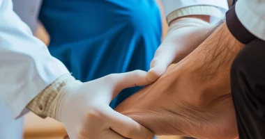
Foreign bodies in the knee
Definition
Foreign (free) bodies in the knee are intra-articular bodies that are partially or completely adjacent to the capsule or other structures of the joint.
Etiology
Many causes can contribute to the formation of free bodies. Cartilaginous and bony intra-articular free bodies are most often formed in degenerative dystrophic diseases; chipped articular cartilage in the form of flakes can result from trauma.
In addition, free bodies are fragments of menisci and cruciate ligaments when they are torn, fibrotic villi of the synovial membrane (from the area of the infrapatellar fold), and tumor fragments.
Less frequently, the source of free bodies is free fixators, such as staples, screws, and remnants of bone cement after endoprosthetic joint replacement. Foreign objects (needles, metal particles, pebbles) rarely become intra-articular free bodies.
Clinical picture
Typically, patients complain of occasional joint locking. In some cases, the main complaints are pain, recurrent synovitis, or painful joint locking associated with joint instability.
In addition, the range of motion may be limited. Large free bodies in the anterior regions of the joint may cause a deficit of extension, while free bodies in the posterior regions cause a limitation of flexion.
Diagnosis
There are no specific clinical tests to diagnose loose intra-articular bodies. Very large loose bodies are easily palpable, and the physician or patient can move them back and forth under the skin. Crunching and clicking noises in the joint indicate severe cartilage damage due to intermittent locking of an unfixed loose body.
Only at least partially calcified free bodies are detected on radiographic examination. Free bodies entirely composed of cartilage are the most common and indistinguishable on radiographs. In all cases, radiographs should be taken in two projections. A Rosenberg projection (anteroposterior projection in 45° knee flexion) is recommended to evaluate the intermuscular notch precisely.
Another option for this purpose is the Frick tunnel projection. In some cases, additional oblique (30°, 45°) or functional projections are required. The maximum flexion projection provides the clearest view of the anterior intercondylar region to confirm or exclude the presence of free bodies, ossifications, or marginal osteophytes.
Errors may occur in the evaluation of radiographs, the most common causes of which are:
- Fabella is a frequently occurring small sesamoid bone localized in the area of attachment of the lateral head of the biceps muscle. In severe cases of osteoarthritis, Fabella may be identified on radiographs as an irregularly shaped mass with indistinct contours.
- Medial patellar facet. Areas of ossification and calcification of the medial patellar retinaculum may appear on radiographs after patellar dislocation. In tangential projections, they acquire a rounded appearance, which makes it possible to suspect intraarticular free bodies. However, free bodies are rarely detected during arthroscopy in this area because ossifications and calcifications are almost always located subcutaneously and inaccessible for arthroscopy.
MRI may be helpful in clarifying the cause of unexplained jamming in the joint and the localization of intra-articular free bodies. However, it should be noted that MRI data are of little use for treatment planning, as arthroscopic removal is indicated if a free body is detected on tomograms. If there are no signs of free bodies, arthroscopy remains the only possible method for further diagnosis and treatment.
Arthroscopic findings
The most common localization.
Large free bodies are localized in areas of the joint that are large enough for them. Small free bodies can easily migrate into tight, hard-to-reach areas of the joint where they are difficult to detect. Migration most often occurs through the hiatus popliteus and through the submandibular spaces, so in cases where a free body is suspected, the lateral and medial menisci should be elevated from the tibial plateau to view the submandibular space. It is sometimes convenient to elevate the menisci with an irrigation cannula. The fluid flow attracts the loose bodies to the opening of the cannula, and they are often evacuated from the joint cavity directly.
Size.
The diameter of free bodies can vary from a few millimeters to several centimeters. Estimating their size is essential for treatment planning.
Large free bodies (greater than two centimeters in diameter) rarely cause joint blockages. They are the most frequent cause of chronic synovitis or limitation of joint mobility, usually intermittent. On the other hand, small and medium-sized free bodies are more likely to migrate into the joint gap, causing episodes of acute joint jamming.
Shape.
Most loose bodies at arthroscopy appear as rounded masses with a pale coloration. The shape and color may indicate their cartilaginous or bony-cartilaginous origin. Free bodies with sharp edges are formed in fresh cartilaginous or cartilaginous fractures and are detected on arthroscopy performed in the first few days after injury. Free bodies with sharp edges, which have been in the joint for a long time, acquire a rounded shape, which occurs not as a result of friction during movement in the joint but as a result of increased activity of fibroblasts, which fill the irregularities of the surface of sharp-edged fragments, forming a rounded free body. Due to the deposition of calcium salts, the free body may increase in size or become partially calcified.
Consistency.
Huge free bodies that appear solid at first glance are often quite friable when palpated with a feeler or grasped with arthroscopic forceps. A homogeneous appearance does not always indicate a dense consistency, and therefore, the instrument used to grasp the free body for removal should be placed with extreme care to avoid the formation of many small fragments of the collapsed body.
Intra-articular loose bodies with a rubber-like consistency are formed from fibrous villi of the synovial membrane, meniscus fragments, or cruciate ligament fibers.
Fixed or unfixed free bodies.
The degree of fixation of the free bodies can be determined by palpation. In fixed free bodies, the attachment area should be examined and evaluated. Sometimes, a flat free body has a large area of synovial contact and may be almost entirely immersed in the synovial membrane. A narrow tissue bridge attaches to the fixed free body in other cases.
The origin of the free body.
The source of free bodies should be determined as a matter of course, as this is of great importance for further treatment and the prognosis of the disease. Suppose the loose body results from a cartilage fracture or Koenig’s disease. In that case, treatment measures should include treating the cartilage surface to induce fibrous growth and even cartilage transplantation.
Secondary injuries.
It is critical to identify any secondary changes caused by free-body impingement. Cartilaginous fractures of the femoral condyles with an anteroposterior fracture line are not uncommon. Free bodies can also cause cartilaginous fractures with exposure of the subchondral bone.
Treatment
In patients with blockages and jamming in the joint, conservative tactics should not be used, as they probably already have a rather deep cartilage lesion. Surgical removal of loose bodies is the method of choice.
Once the free body has been localized, it is necessary to determine whether it is fixed. Fixed free bodies can cause joint blockages by slipping into the intermuscular space. The operation aims to remove the free body, identify the cause of its formation, and treat secondary injuries.
All these treatment options are available in more than 790 hospitals worldwide (https://doctor.global/results/diseases/foreign-bodies-in-the-knee). For example, Arthroscopic knee debridement can be done in these countries at following approximate prices:
Turkey$3.9 K in 26 clinics
Germany$13.2 K in 43 clinics
China$13.6 K in 8 clinics
Israel$18.0 K in 16 clinics
United States$22.9 K in 17 clinics.
Postoperative management
If the intervention consisted only of removing a fixed or non-fixed free body, in the absence of pain syndrome or swelling, the patient was recommended dosed loads (50% to full by the third day of the postoperative period).
Suture removal is performed from the fourth to the sixth day of the postoperative period. Lymphatic drainage massage is indicated if there is effusion in the joint. Otherwise, the postoperative rehabilitation protocol depends on the associated intra-articular injuries.