
Mitral valve insufficiency
What’s that?
Mitral valve insufficiency, or mitral regurgitation (MR), is a congenital or acquired defect of the mitral valve, which results in the reverse blood flow from the left ventricle to the left atrium during heart function. Pathology takes the first place among all valve defects of the heart. This disease is quite rare in isolation. In 50% of cases, it develops in the structure of combined and concomitant heart defects.
About the disease
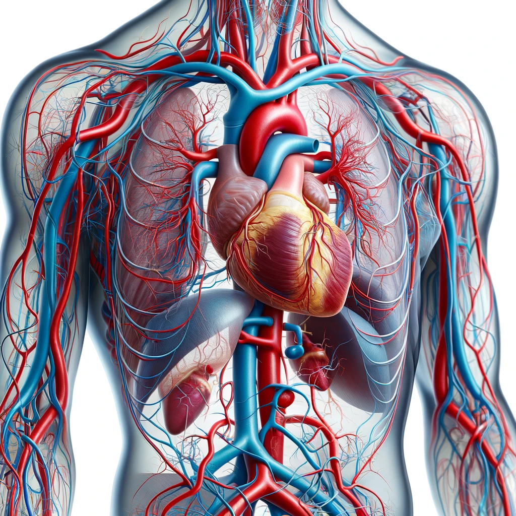
The mitral (bicuspid) valve is located between the left ventricle and atrium and is responsible for the proper flow of oxygenated arterial blood. Usually, it closes as soon as blood enters the ventricle, then the heart muscle pushes it to the aorta and all organs except the lungs. In mitral insufficiency, the valve loses its ability to close tightly, which causes mitral regurgitation, i.e., partial blood flow in the opposite direction.
Types of mitral valve insufficiency
There are several types of classification of mitral valve insufficiency based on various factors. This pathology can also beacute and chronic.
Heart defects are categorized into two types according to their origin:
- Congenital or primary: these are genetic abnormalities of chord structure leading to flap prolapse;
- Acquired or secondary; this includes mitral valve deterioration resulting from other diseases or injuries.
Depending on the provoking cause of the pathology, mitral insufficiency is subdivided into:
- ischemic after a myocardial infarction;
- organic, caused by structural changes in the valve itself or in the chordae tendineae that hold it together;
- functional, arising against the background of left ventricular pathologies.
In the clinical course of the disease, three stages are distinguished:
- Compensated. Changes in the mitral valve are insignificant. Regurgitation is 20-25% of the blood volume entering the aorta at each heart contraction (systolic volume).
- Subcompensated. Blood return to the left atrium is 25-50% of its systolic volume, and the patient shows the first signs of cardiac pathology.
- Decompensated. Regurgitation is 50-90% of the systolic volume, and global heart failure develops, which often leads to pulmonary edema.
Symptoms of mitral valve insufficiency
There are no clinical signs at the pathological process’s initial stage. The development of MR can be detected only during cardiac ultrasound examination. The prognosis depends on the hole size that allows blood to flow back into the left atrium. In several patients, signs of ischemia (lack of oxygen) of the myocardium and other organs appear, and stasis occurs in the pulmonary vessels. In this case, signs of mitral insufficiency usually include:
- atrial fibrillation, increased heart rate;
- fatigue when performing everyday activities;
- marked soreness on the left side of the chest;
- sensation of shortness of breath, first on exertion and then at rest;
- cough, increasing in the supine position and accompanied by the appearance of sputum with an admixture of blood;
- swelling of the legs (mild swelling accompanied by pallor and impaired skin elasticity).
On examination of the patient, several objective signs of mitral valve insufficiency are noted:
- bulging neck veins;
- heart murmurs;
- acrocyanosis (blue skin color of the tips of the ears, nose, and limbs) against the background of general pallor of the skin, etc.
Causes of mitral insufficiency
The development of congenital heart disease is associated with the impact of negative factors on the fetus during intrauterine development:
- infection with rubella, herpes, etc;
- exposure to radiation or chemicals;
- intoxication with alcohol, tobacco smoke, drugs, and medicines.
The causes of hereditary (genetic) mitral valve insufficiency are related to chromosomal abnormalities in the family history. The development of acquired mitral regurgitation is most often provoked by:
- Acute rheumatoid arthritis is the most common cause of developing MR;
- Ischemic heart disease, the second of the main causes leading to the development of mitral regurgitation;
- Infective endocarditis;
- Systemic diseases (SLE and others);
- Cardiac tumors (more often atrial myxoma);
- Amyloidosis, sarcoidosis.
The risk group for developing this pathology includes patients with prosthetic valves. MR develops in case of loss of stability of the mechanical structure or degeneration of the biological prosthesis.
Diagnosis of mitral insufficiency
When a patient with a suspicion of mitral insufficiency is assigned a complete diagnostic study, the primary methods that allow the identify the development of the pathological process and the correct diagnosis are:
- Physical examination – the specialist measures blood pressure, listens to the pulse, listens to the lungs and systolic murmurs.
- Echocardiography is an ultrasound examination to evaluate the structure, condition, and size of the heart muscle, large vessels, and valves.
- Electrocardiography – records the bioelectrical potentials of the heart. At the terminal stage of the pathology, arrhythmias are usually recorded.
- X-ray – shows indirect signs: enlargement of the heart muscle—symptoms of pulmonary congestion.
Laboratory diagnosis aims to identify the causes of pathology and assess the condition of other organs. Differential diagnosis of mitral valve insufficiency allows us to distinguish this pathology from other heart defects, severe myocarditis, or dilated cardiomyopathy.
Treatment of mitral insufficiency
Therapeutic measures in this pathology are aimed at:
- symptoms management;
- reducing the risk of possible complications;
- increasing the patient’s life expectancy.
Conservative treatment
Conservative treatment of mitral valve insufficiency can be performed only in the early stages of the disease or in preparation for surgery. No particular therapy is required in asymptomatic chronic mild form of MR.
Drug therapy for this pathology may include the prescription of the following groups of drugs to the patient:
- antiarrhythmic drugs that regulate heart rate;
- antiplatelet drugs, medications that prevent platelets from sticking together;
- cardiac glycosides that improve myocardial tone;
- diuretics to reduce swelling in the extremities.
To stabilize hemodynamics, the patient may be given intra-aortic balloon counterpulsation (pumping blood into the aorta with a particular medical pump).
Surgical treatment
Surgical interventions are indicated in II-III degree insufficiency. Surgery aims to restore the mitral valve’s closure function. In most cases, prosthetic replacement of the mitral valve with an artificial or biological prosthesis is performed.
In the absence of pronounced changes, it is possible to perform valve-preserving operations:
- narrowing of the annulus fibrosus;
- replacement of a valve leaflet;
- mitral valve repair, etc.
The correction is carried out after the heart is disconnected from circulation. A heart-lung machine performs its function during surgical intervention. Thanks to surgical intervention, achieving good results without complications is possible.
All these surgical options are performed in more than 680 hospitals worldwide (https://doctor.global/results/diseases/mitral-valve-insufficiency). For example, Heart valve repair can be performed in 29 clinics across Turkey for an approximate price of $8.4 K (https://doctor.global/results/asia/turkey/all-cities/all-specializations/procedures/heart-valve-repair).
Prevention of mitral valve insufficiency
People with congenital mitral insufficiency should undergo screening examinations by a cardiologist and a cardiac surgeon. Prevention of acquired MR consists of preventing diseases that provoke its development. It is essential to treat streptococcal infection timely, because it often causes complications mitral regurgitation.
Rehabilitation
The patient spends the first day after surgery in the intensive care unit, and then, in case of sound postoperative period, patient is transferred to a general ward. Rehabilitation in the hospital lasts 7-10 days. At discharge, the attending physician gives recommendations on lifestyle for 6-12 months until complete recovery. They are individualized for each person and depend on age, condition, and extent of the surgical intervention. For example, lifelong intake of medications, compliance with work and rest, proper nutrition, restriction of bad habits, dosed physical activity, etc.
