Osteophytes
What’s that?
Osteophytes (exostoses) are growths of bone tissue that form on the surface of joints due to arthritis, arthrosis, other degenerative or inflammatory diseases, and trauma to the musculoskeletal system. In rare cases, such growths become an independent pathology. The main danger lies in the growth of osteophytes: their increase leads to displacement of tissues and mechanical damage to internal structures, which can eventually result in complete immobility of joints and disability.
About the disease
Intensive activity, age-related thinning of tissues, and inflammatory and other pathological processes – all lead to cartilage atrophy, which causes the articular surfaces of bones to rub against each other and destroy. To prevent such changes, the body accelerates the growth of bone cells, which causes outgrowths to form. They reduce the amplitude of movement of the damaged areas, thereby reducing the load. The formation of osteophytes can be called a protective reaction of the body.
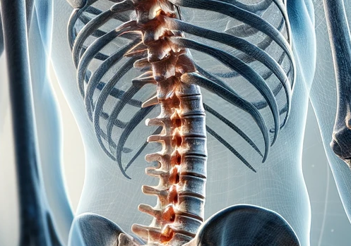
Most often, bone growths occur in large joints: hip and knee joints, spinal column, heel bone, etc. Osteophytes are visible on radiological images: in the initial stages of development, they first look like small spots on the surface of the bone and, with extensive growths, visually resemble spikes.
Types
Specialists, taking into account clinical symptoms, volume, and type of bone tissue changes, distinguish four main stages in the development of osteophytes:
- At the first stage, there are no external signs, no pathologic foci of ossification can be seen on radiography;
- In the second stage, the patient begins to experience pain in the affected area after physical activity, sharp turns and movements, and single growths and narrowing of the articular gap become visible on the scans;
- The third stage is accompanied by pronounced symptoms (pain, restriction of motor activity, swelling), while on X-ray images, the distance between the joint elements is significantly narrowed, and there are signs of multiple exostoses;
- In the fourth stage, the joint is severely limited in movement, the patient experiences constant pain and stiffness, and the joint gap is absent in images, but there are pronounced signs of multiple osteophytes.
Given the localization, exostoses can be vertebral, hip, cervical, knee, etc.
Symptoms
Clinical signs of osteophytes depend on the localization of the pathological process and the stage of development of outgrowths. The key ones are:
- dull, aching pain in the area of the affected joints, which intensifies after physical activity, during movement, turning;
- muscle weakness;
- rapid physical fatigue;
- numbness, tingling sensation in the area of localization of exostoses;
- swelling of soft tissue in the injured area.
The pain syndrome is more pronounced if the osteophytes are located on the sole of the foot. This condition is called heel spur (plantar fasciitis) and is accompanied by severe pain when walking.
The syndrome is generally characterized by subsiding discomfort and soreness at rest.
With multiple osteophytes that damage tissue and compress nerve fibers, symptoms may also include:
- decreased sensation in the lower and upper extremities;
- urinary disturbances;
- diarrhea;
- stiffness when moving, especially after staying in one position for a long time (after sleeping, a long ride in a motor vehicle, etc.).
In the future, the patient will find it increasingly difficult to move, his gait will change, movements will become slower and more uncertain, and their amplitude will decrease.
Reasons
Any inflammatory diseases of joints and bone structures and degenerative and age-related processes are considered risk factors. The most common causes of osteophytes are:
- different types of spinal curvature (kyphosis, scoliosis);
- obesity, which repeatedly increases the load on joints and cartilage;
- wearing tight, small, and uncomfortable shoes;
- endocrine, autoimmune diseases;
- presence of cancer in the patient’s history;
- bone and joint injuries;
- excessive physical exertion, which the patient experiences regularly.
Factors predisposing to the formation of exostoses also include:
- sedentary lifestyle;
- smoking, drinking alcohol;
- chronic stress conditions;
- intense exercise in certain sports (running, walking, cycling, contact games, etc.);
- age over 50.
The chances of developing diseases accompanied by the formation of exostoses are higher in people who are engaged in teaching and have to spend a lot of time standing, walking, or carrying heavy loads (couriers, letter carriers, movers, etc.).
Diagnosis
At the initial meeting, the doctor collects an anamnesis, conducts an external assessment of the musculoskeletal system, and performs palpation of the affected joints. A general neurological examination is prescribed to determine the negative impact of pathology on the CNS.
The set of instrumental studies includes:
- X-ray of the affected area in several projections, allowing to determine the type and localization of osteophytes;
- ultrasound of soft tissues and joints;
- electroneuromyography – a particular procedure that helps to assess the functionality of the nervous system in the peripheral areas;
- computer and magnetic resonance imaging allow us to determine the stage of the pathological process, identify the features of exostoses, detail, and study all the necessary structures.
In addition, several laboratory tests are performed. If the patient has systemic diseases, various specialists are involved in consultations: endocrinologists, traumatologists, rheumatologists, etc.
Treatment
Treatment tactics for diseases in which osteophytes are formed depend on the age of the patient, the condition of the patient’s bones and joints, clinical symptoms, the presence of concomitant pathologies, and a number of other parameters.
There are two main approaches to therapy: conservative and radical. Conservative modalities include:
- medication;
- physical therapy and gymnastics;
- nutritional and lifestyle adjustments;
- obesity treatment.
The list of medications used in the therapy of diseases of bone and cartilage structures may include:
- anti-inflammatory drugs and painkillers;
- chondroprotective drugs;
- antibiotics for active infection.
The patient needs to give up bad habits, have a proper and nutritious diet, lose weight, and give the body a little exercise every day.
If conservative measures have no effect, the patient’s condition is severe, and there is a risk of complete immobilization of the joints, surgical treatment may be prescribed. It consists of resection (excision) of osteophytes, sometimes together with an area of damaged bone and cartilage structures.
In cases of extensive inflammation, arthroscopy is performed, a procedure that allows drugs to be delivered directly to the focus of inflammation. During arthroscopy, surgeons can remove minor exostoses and perform several other manipulations.
All these treatment options are available in more than 950 hospitals worldwide (https://doctor.global/results/diseases/osteophytes). For example, ankle osteophyte removal is performed in 45 clinics across Germany for an approximate price of $4,970 (https://doctor.global/results/europe/germany/all-cities/all-specializations/procedures/ankle-osteophyte-removal).
Prevention
To minimize the risk of developing diseases in which exostoses form, it is desirable:
- adhere to an active lifestyle, regularly giving the joints adequate load (do exercises, walk in the fresh air, exercise, etc.);
- avoid wearing tight and narrow shoes;
- exercise regularly, especially weightlifting, contact sports, martial arts, and wear protective equipment to avoid injury;
- do not overeat; control your body weight by keeping your weight within your BMI.
People whose occupation involves extended periods of sitting should regularly (at least once every 1.5-2 hours) get up and do a light warm-up. For those who have to walk or stand a lot, it is desirable to carefully choose shoes and organize their work regime to minimize the load on the spine and legs.
Rehabilitation
After surgical treatment, recovery takes 1 to 6 months, depending on the type of surgery performed, the patient’s initial condition, and other essential criteria.
During rehabilitation, it is essential to:
- To keep as much rest as possible;
- Do not come into contact with people who have had infectious diseases in the last 2-4 weeks;
- To stick to a gentle diet;
- Carefully follow the doctor’s orders and prescriptions.
It is essential to understand that if the patient does not take care of his health, does not fight diseases or bad habits, is overweight, and continues to test the joints for strength, even surgical treatment will not eliminate the repeated formation of osteophytes.


