
Portal hypertension
What’s that?
Portal hypertension is a pathologic condition that is accompanied by an increase in pressure in the portal vein system.
About the disease
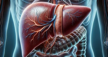
The portal vein is a collector of blood from venous vessels collecting blood from unpaired organs located intraperitoneally. These include the small and large intestine, stomach, pancreas, gallbladder, and spleen. The portal vein penetrates the liver in the transverse fissure region, forming a robust venous network in the parenchyma. The causes of portal hypertension are diverse, but 85% of cases are caused by cirrhotic transformation of the liver.
Increased pressure in the portal vein leads to the opening of shunts with other venous vessels. Over time, this compensatory response, initially aimed at reducing hypertension, leads to pathologic changes. Thus, the most dangerous and common complication of portal hypertension syndrome is the expansion of esophageal and gastric veins with subsequent bleeding from them. The insidiousness of such a condition is that it is prone to recurrence. Within a year, bleeding recurs in 28-70% of cases and within two years – 80-90% of observations. In every third patient, recurrent bleeding is resistant to conservative treatment, and severe blood loss leads to a sharp decompensation of liver function.
Diagnosis of portal hypertension is based on clinical data, laboratory, and instrumental studies. The comprehensive examination allows us to determine the localization of the pathological process, the severity of hypertension in the portal vein, and the risk of complications.
In the first stage, conservative treatment of portal hypertension is carried out. If the pathological process progresses, the question of surgical treatment is decided. Surgery may also be indicated in case of bleeding from pathologically altered esophageal varicose veins.
Types of portal hypertension
In gastroenterology, the following pathogenetic types of portal hypertension are distinguished:
- suprahepatic type;
- intrahepatic type;
- extrahepatic type;
- mixed type.
Symptoms of portal hypertension
The clinical form determines the symptoms of portal hypertension. Most often, the patient is concerned with manifestations of dyspepsia syndrome:
- lack of appetite;
- nausea;
- flatulence;
- diarrhea;
- general weakness;
- poor tolerance for physical activity;
- the feeling of heaviness and pain in the subcostal region, not only on the right but also on the left.
According to ultrasound findings, signs of enlargement of the liver and spleen are detected.
The etiology of the disease (intrahepatic or extrahepatic origin of portal hypertension) affects the peculiarities of symptomatology.
- The extrahepatic form is characterized by an acute onset, a pronounced pain syndrome in the epigastrium and right subcostal region, a sharp increase in the size of the liver and spleen, and increasing ascites accompanied by fever.
- The leading symptoms of the intrahepatic form are jaundice, large size of the liver and spleen, the presence of hepatic signs (palmar redness, vascular elevations on the skin, called telangiectasia), ascites, the formation of visible venous anastomoses on the anterior wall of the abdomen, the presence of edema-ascites syndrome and signs of hypersplenism (due to increased spleen function, platelets and red blood cells are destroyed, which is manifested by bleeding of various localizations and anemia).
The intrahepatic form of portal hypertension is most often manifested by bleeding from esophageal veins. The following symptoms characterize this complication:
- vomiting like “coffee ground” (the brown color is due to oxidation of hemoglobin);
- black liquid stool (black color is due to hemosiderin formed during the chemical transformation of hemoglobin).
In portal hypertension, bile and lymph formation are often impaired, which contributes to the deterioration of the bacterial-filtering function of the lymphatic system. In turn, this creates conditions for endogenous intoxication due to vascular shunts and inadequate function of stellate reticular endotheliocytes, which normally inactivate enterogenic toxins.
Portal hypertension is insidious, with the development of bleeding from gastric and esophageal veins. A combination of several interrelated causes plays a role in the pathogenesis of this condition:
- the increase in portal pressure, the severity of portal stasis;
- morphologic restructuring of the vessels of the portal basin, the formation of esophageal-gastric varicose veins;
- the presence of inflammatory changes in the mucosa of the esophagus and cardiac gastric region, the severity of which depends on the degree of tissue nutrition disorders, weakening of esophageal motility, the existence of gastroesophageal reflux;
- violation of blood coagulation processes, which may be accompanied by thrombotic and hemorrhagic manifestations at the level of macro- and microcirculation.
Diagnosis of portal hypertension
The following tests are used for diagnosis when portal hypertension is suspected:
- general clinical blood test;
- blood biochemistry test;
- analysis of the blood coagulation system, including evaluation of INR and D-dimer;
- determination of markers of viral and autoimmune hepatitis;
- determination of indicators of iron and copper metabolism;
- ultrasound scanning of the liver and abdominal organs with an assessment of the vascular bed;
- endoscopic examination of the stomach and others.
On ultrasound, portal hypertension is indicated by the following signs:
- increased diameter and tortuosity of the portal vein (more than 13 mm);
- increase in the lumen of the splenic vein (more than 8 mm);
- dilation of the superior mesenteric vein;
- exceeding the normal size of the liver and spleen;
- the presence of vascular connections between the portal vein system and the inferior vena cava;
- detection of signs of ascites in the abdominal cavity.
The gold standard for diagnosing portal hypertension is the venous pressure measurement in the portal vein. It is an invasive study performed using a cardiac catheter inserted through the ulnar vein. An increase in the difference in measurements of more than ten mmHg confirms the diagnosis of portal hypertension. However, given the invasiveness of the study, ultrasound with color Doppler mapping is currently used as a screening diagnosis.
The diagnostic search program may include computed tomography and magnetic resonance imaging with contrast. These methods allow us to obtain a clear image of parenchymatous organs, large vessels, retroperitoneal space, and other structures, which makes it possible to identify the cause of portal hypertension. Clear visualization of vessels helps to determine the level of blood flow block in the portal vein and the presence and severity of collateral blood flow.
Liver biopsy is the gold standard in diagnosing fibrosis as a cause of portal hypertension. When performing a biopsy with subsequent histologic examination, the doctor obtains a morphologic picture characteristic of the disease that led to impaired blood circulation in the portal vein system. If there are no pathologic changes in the biopsy, it is considered that the cause is associated with the pre-hepatic block. To determine the stage of fibrosis with a high degree of reliability of the result, a modern non-invasive method – liver fibroscan – is used.
An essential diagnostic element in determining the source of bleeding from varicose veins is esophagogastroscopy, performed in the first hours after the patient’s admission to the hospital. When performing endoscopy, there is a favorable opportunity for emergency sclerotherapy in patients with ongoing and spontaneously stopped bleeding.
Treatment of portal hypertension
According to clinical recommendations, conservative approaches to treatment can be successfully applied in case of an insignificant pressure increase in the portal vein. In case of progression of the pathological process, surgical intervention is performed.
Conservative treatment
The main directions of conservative therapy are:
- prevention of bleeding from dilated esophageal veins with beta-blockers and angiotensin-converting enzyme inhibitors;
- control of ascitic syndrome with diuretics, changes in dietary habits (decreasing salt intake, increasing the proportion of protein products);
- therapy for a background disease.
Surgical treatment of portal hypertension
Surgical treatment of portal hypertension is palliative and aimed at unloading the portal vein. The only method that allows the problem to be solved radically is liver transplantation.
Laparocentesis is used quite successfully to evacuate ascitic fluid from the peritoneal cavity. It is one of the most common procedures in surgery.
Types of surgery for portal hypertension
The main types of surgery for portal hypertension are:
- creation of new outflow routes from the portal vein, for example, its anastomosis with the inferior vena cava (portocaval anastomosis);
- interruption of the connection between the venous system of the gastroesophageal segment and the portal system (removal of a part of the stomach);
- X-ray-guided endovascular interventions (e.g., intrahepatic portosystemic shunt);
- reduction of blood flow into the portal vein (for this purpose, the spleen is removed).
Splenectomy surgery is not a method of reducing pressure in the portal vein. Still, it has its indications, such as the presence of arteriovenous fistulas and extrahepatic form of portal hypertension (so-called left-sided portal hypertension in pancreatic diseases).
All these treatment options are available in more than 680 hospitals worldwide (https://doctor.global/results/diseases/portal-hypertension). For example, portocaval anastomosis can be performed in 38 clinics across Germany (https://doctor.global/results/europe/germany/all-cities/all-specializations/procedures/portocaval-anastomosis).
Prevention
Prevention of portal hypertension is primarily aimed at preventing viral hepatitis. For this purpose, vaccination against hepatitis B is carried out, and it is recommended to observe safety measures against other hepatotropic viruses, which can be transmitted by sexual contact or through infected blood. In the presence of background pathology, adequate correction is indicated, which reduces the risk of portal hypertension.
Rehabilitation
After surgery, it is essential to adhere to a diet and eat small portions of food, but often (5-7 times a day). Reducing salt intake and increasing the proportion of protein products in the diet is essential. After surgery, early activation is indicated.