Shoulder bursitis
What’s that?
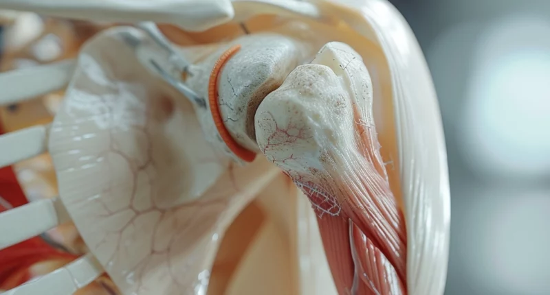
Bursitis of the shoulder joint is an inflammatory lesion of the synovial bag of the corresponding joint.
About the disease
The articular pouch is a narrow cavity filled with synovial fluid. Bursae are formed in places where the bony structures of the joint approach the skin, thus performing a delimiting and smoothing function. When the inflammatory process develops, the synovial cover begins to produce a large amount of fluid while its composition changes. It becomes the morphological substrate for the development of the corresponding symptoms of bursitis of the shoulder joint.
The disease can have an infectious and non-infectious nature. The main manifestations of bursitis are pain in the shoulder joint, limited mobility, and skin swelling. In case of impingement of nerve trunks passing nearby, numbness and weakness of muscle strength may develop.
The inflammatory process most commonly involves the subacromial and subdeltoid pouches. Sluggish, non-infectious lesions of the shoulder joint bags are most often found in young people who are at risk of traumatization of soft tissues, for example, with heavy sports activity and non-compliance with the technique of exercise. Given that the main factor of the disease is chronic traumatization, then the risk group includes miners, loaders, dentists, raters, etc. From what else can bursitis happen? Rheumatologic bursitis, associated with autoimmune processes, is allocated in a separate category.
The diagnosis is established based on clinical symptoms and the data of additional examination, in particular imaging methods (radiography, ultrasound scanning, computer or magnetic resonance imaging). Laboratory tests allow to establish the activity of the inflammatory process.
The list of applied methods of treatment of inflammatory lesions of the bursae of the shoulder joint is quite extensive and is represented by various options for local and general treatment. For rapid pain relief, local therapy in the form of therapeutic injections of analgesics in combination with glucocorticoids is performed.
Types
Bursitis is classified into two types according to its course:
- acute;
- chronic.
By localization, the pathological process may be as follows:
- subacromial;
- subdeltoid;
- subcoracoid.
This localization is determined by which pouch is involved in the inflammatory process. If the inflammation affects the area under the deltoid muscle, it is called subdeltoid bursitis. If the pouch near the acromial process of the scapula becomes inflamed, it is subacromial bursitis, and if it is in the area of the coracoid process, it is subcoracoid bursitis.
Symptoms of shoulder bursitis
The main clinical signs of bursitis may include:
- moderately pronounced pain sensations in the joint area;
- limitation of mobility in the shoulder joint (due to pain);
- redness of the skin over the articulation (hyperemia is moderately pronounced);
- swelling of the skin over the shoulder joint.
As a rule, the patient’s general condition in bursitis does not suffer. When the purulent process develops in the articular bag, body temperature may increase slightly.
Pain and local increase in skin temperature are determined when palpating the affected joint. When extending and flexing the joint (actively and passively), there is an increase in pain.
Timely initiation of treatment contributes to the acute process ending with recovery. If therapy is not carried out, the disease becomes chronic. Pain, in this case, becomes less pronounced, swelling decreases, hyperemia passes, and a local tumor of soft elastic consistency can be determined, corresponding to the inflamed bursa. Chronic bursitis is characterized by the appearance of unpleasant sensations during movement in the joint, as well as the limitation of the amplitude of these movements. The patient may be bothered by paresthesia, including numbness of the hand—the muscles surrounding the joint reflexively spasm.
In the purulent form of bursitis, clinical signs are more vivid. The patient is bothered by intense pain in the joint, rasping or twitching character, increased body temperature, and other manifestations of intoxication syndrome. The inflamed joint is hot, swollen, and painful to the touch. In the general clinical blood test, the level of leukocytes is increased, erythrocyte sedimentation is accelerated, and there is an increase in the level of neutrophils.
Causes of shoulder bursitis
The causes of shoulder bursitis can be as follows:
- Constant exposure of the joint to ultra-strong mechanical loads that exceed the limit of its strength;
- Direct trauma to the joint, e.g., due to a blow, sprained ligaments;
- Metabolic disorders – uric acid salts in gout can deposit in the joint capsule, leading to the development of bursitis;
- Autoimmune processes, in which the cells of the immune system begin to attack the soft tissue structures of the joint;
- Direct penetration of microorganisms into the joint cavity during open wounds, purulent skin lesions (pimples, abscesses), as well as due to the spread of pathogens with blood and lymphatic flow from foci of chronic infection (pyelonephritis, tonsillitis, lymphadenitis, etc.).
When the synovial pouch becomes inflamed, the synthesis and viscosity of synovial fluid increases, and the bursa’s synovial membrane thickens. From a practical point of view, it is essential to distinguish between infectious bursitis, caused by microorganisms, and aseptic bursitis, caused by traumatization, urate deposition, or the development of autoimmune reactions. In the case of the infectious nature of the disease, the main causative agents may be staphylococci and streptococci, pseudomonas bacillus, and other pathogens.
Diagnosis
To visualize inflammatory changes in the joint, two screening tests are performed – radiography and ultrasound scanning. Radiography allows the assessment of the condition of bony structures and the detection of calcium salt deposits in the joint bag, which usually indicate a chronic course of the disease (calculous or stony bursitis). Ultrasound scans are more informative for determining the condition of the soft tissue structures of the joint. Ultrasound helps to determine which shoulder bag is inflamed, how active the inflammation is, and the approximate volume of exudate in the synovial bag.
In complex clinical cases, CT or MRI is indicated. These studies allow obtaining layer-by-layer scans of the studied area with a 2-3 mm step. A 3D model can be built based on the images to visualize the nature and extent of the detected changes.
If purulent bursitis is suspected, a general clinical blood test is performed, which reveals signs of bacterial inflammation. If it is necessary to exclude the rheumatologic nature of the disease, rheumatological tests (antistreptolysin-O, rheumatoid factor, etc.) are indicated. Biochemical blood tests can determine uric acid levels if gout and associated bursitis are suspected.
Treatment of shoulder bursitis
The treatment program for bursitis is determined by the severity and nature of the inflammatory process, the severity of the pain syndrome, and the presence of concomitant pathology. In most cases, complex conservative therapy is sufficient. However, surgical intervention is indicated in the case of a purulent process.
Conservative treatment
Complex conservative therapy of bursitis consists of the following measures:
- medications;
- physical therapy;
- massage.
Nonsteroidal anti-inflammatory drugs of local or systemic application are used to control pain. In severe cases, injections of corticosteroids into the joint cavity are indicated. A supraspinous nerve block is performed if this measure fails to achieve therapeutically significant pain relief.
Physical procedures are widely used in the complex treatment of shoulder joint bursitis. The fastest and most effective action is recognized as extracorporeal shockwave therapy. The therapeutic effect of the shock wave is as follows: high-energy shockwave vibration briefly affects the focus of the disease, causes a decrease in pain syndrome, improves local blood circulation, and reduces inflammation of fibrotic foci.
Surgical treatment
Surgery for purulent bursitis consists of opening the inflamed articular bag using endoscopic instruments, evacuating pus and destructively altered tissues, washing the bag with antiseptic solutions, and applying a drain. The latter provides an unimpeded outflow of pathologic secretion. In addition, injections of corticosteroid drugs inside the joint can also be used.
All these treatment options are available in more than 820 hospitals worldwide (https://doctor.global/results/diseases/shoulder-bursitis). For example, shoulder arthroscopy can be performed in 14 clinics across Israel for an approximate price of $6.5 K (https://doctor.global/results/asia/israel/all-cities/all-specializations/procedures/shoulder-arthroscopy).
Prevention
To reduce the risk of shoulder bursitis, it is essential:
- observe occupational hygiene;
- adhere to proper exercise technique, apply taping;
- treat infectious processes promptly so as not to contribute to the formation of foci of chronic infection in the body.
Rehabilitation
After the surgical opening of the inflamed bursa, it is essential to provide functional rest to the operated arm. For this purpose, special supportive dressings are used. After wound healing, a course of massage and physiotherapy is performed.


