
Skin cancer
What’s that?
Skin cancer is a malignant epithelial tumor that affects the dermis and is prone to metastasis.
What skin cancer means
Skin cancer accounts for about 10% of all cancers. Elderly patients are at risk, and the likelihood of skin cancer is the same for men and women. It is proved that people with light skin type who live in hot countries or high mountains, i.e., in those regions where solar activity is maximum, have higher cancer risks. Patients who spend long periods outdoors due to their work are also at risk.
Skin cancer gradually spreads deep into the tissue and causes tissue lysis. If the tumor process develops on the head and face, it can affect the sensory organs (ears, eyes, nasal cavity, and sinuses) and quickly reach the brain. Tumors can be localized on any body part – legs, thigh, arms, hands, face, back, fingers, temple, forehead, elbow, lips, chest, neck, heel, cheek, stomach, and foot.
The spread of cancer cells through blood vessels can lead to the formation of distant foci of dropout. They can be localized in various internal organs. The zone of metastatic lesions can capture the digestive tract, bones, urinary organs, mammary glands, and other organs.
Treatment of skin cancer is based on the removal of the malignant tumor. Radiation therapy or chemotherapy may be performed as indicated.
Types of skin cancer
According to the histologic picture, the following types of skin cancer are distinguished:
- squamous cell variant – the precursor is atypical cells of the epidermal layer of the skin;
- Basal cell variant (basalioma) – occurs due to malignant transformation of epithelial stem cells, which generally provide its renewal and regeneration;
- Adenocarcinoma – a tumor from the category of casuistic, it comes from the cellular structures of sebaceous or sweat glands;
- melanoma – this variant of malignant tumor develops from melanocytes (skin cells containing the pigment melanin).
In clinical practice, there is an established opinion that, given the unique characteristics of melanoma (aggressiveness, early metastasis), it should be singled out in a separate category of malignant epithelial tumors. And when talking about skin cancer, most often, we mean those forms that are non-melanoma.
The clinical diagnosis reflects the stage of the cancer process, which is determined based on the characteristics of the process according to the TNM classification.
T – characteristics of the primary tumor (size and spread to underlying deep tissue);
N – status of the nearest lymph nodes, which is determined by the presence or absence of tumor cells in them;
M – presence of metastasis (spread of malignant cells to distant areas).
The G category also plays a vital role in determining treatment tactics. It reflects the degree of differentiation (maturity) of the tumor cells. The more mature the cells are, the better the prognosis of the disease. Highly differentiated tumors are coded as G1 and undifferentiated tumors as G4.
Symptoms of skin cancer
The histologic variant of the tumor largely determines the symptoms of skin cancer.
Squamous cell skin cancer
This variant of a malignant tumor is characterized by a rapid increase in the neoplasm and a tendency to invade both on the surface of the skin and in-depth. The tumor does not hurt for a long time. The appearance of painful sensations usually indicates the spread of the malignant process to the bones, muscles, or cartilage nearby. In some cases, the pain may not indicate that an infectious inflammation has set in.
Visual signs of squamous cell cancer may include the following elements:
- ulcerative defect;
- a plaque-like mass;
- nodal seal.
Ulcerative defects in squamous cell skin cancer resemble a crater, bordered by dense elevated edges with a steep precipice—the bottom of the epithelial defect with irregularities, covered with dried spongy crusts.
A bright red color characterizes the plaque-like form of skin cancer and has a dense structure and superficial tubercles. It is characterized by rapid growth and arrosion of vessels, which leads to the appearance of bloody discharge. In the presence of such symptoms, it is important not to ignore the problem and consult a dermatologist or oncologist.
The nodular form of squamous cell cancer is characterized by a formation resembling a cauliflower or mushroom appearing on the skin. The color of the tumor is bright red or brown; the structure is very dense. Its surface may be eroded or ulcerated.
Basal cell cancer
Basalioma is characterized by a relatively benign course compared to tumors of squamous cells. Only with the prolonged existence of the disease is the sprouting of the underlying tissues possible, accompanied by painful sensations. Basaliomasrarely and late metastasize, which determines a relatively favorable disease prognosis.
Adenocarcinoma
This variant of malignant tumor most often occurs in areas with a vast number of sweat and sebaceous glands. Therefore, the favorite areas for localization of adenocarcinoma are the armpit, groin, and sub-mammary fold (under the breast).
The onset of the disease is characterized by forming a nodule that rises above or in tone with the skin. This type of skin cancer, which has a very low incidence in the population, is characterized by slow growth.
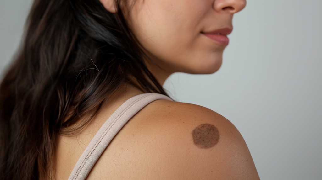
Melanoma
Most often, melanoma appears as a stained tumor. Its hue can be different but within the black-brown or gray range. Extremely rarely, melanoma may have no coloration. This depigmented variant presents the most significant diagnostic difficulties. In the development of melanoma, 2 phases are distinguished:
- Horizontal growth phase, when the tumor spreads quite rapidly over the skin surface;
- The vertical growth phase is when tumor cells spread deep into the dermis.
Causes of skin cancer
The leading cause of malignant transformation of epithelial cells is considered to be prolonged exposure to aggressive doses of ultraviolet light. The role of this causative factor is confirmed by the fact that in almost 90% of cases, cancerous skin tumors develop on the exposed skin, particularly on the face and neck. People with a light skin type are at risk, as its low pigment content does not retain natural ultraviolet light well.
In addition, some chemical substances that are exposed to the skin for a long time are significant factors of carcinogenesis. Thus, tobacco smoke, arsenic compounds, tar substances, fuels, and lubricants pose a cancer risk. In some cases, skin cancer can develop at the site of a burned surface or be a complication of radiation dermatitis, which proves the role of physical factors in skin carcinogenesis. Frequent traumatic injury to scarred areas or benign nevi can also cause skin cancer.
Diagnosis of skin cancer
According to clinical guidelines for skin cancer, patients with suspected skin cancer are consulted by a dermatologist-oncologist. The doctor conducts a visual assessment of the skin, palpation of lymph nodes close to the lesion area, and dermatoscopy. Ultrasound scanning can be performed to identify the invasiveness of the process. Pigmented neoplasms are additionally subjected to SIAscopy, revealing even minimal structure changes.
To make a definitive diagnosis of a cancerous skin tumor allows morphological examination, which can be in 2 variants:
- Cytologic diagnosis involves assessment of the cellular composition of altered tissues; the material is smears taken from the areas of epithelial defect;
- histologic diagnosis – performed on material obtained after removal of skin cancer or by puncture plucking (biopsy). Histologic diagnosis reveals the presence of atypical cells and tissue structure and establishes their origin (squamous, basal, melanocytes) and degree of differentiation.
Skin cancer treatment
The choice of skin cancer treatment is determined according to its histologic type, the extent of the cancer process, and the degree of differentiation of cancer cells. The localization of skin cancer and the patient’s age are also considered. The treatment program consists of removing the malignant tumor and deciding whether radiation therapy is necessary.
Conservative treatment
Skin cancers that cover a small area can be effectively treated with close-focus X-ray therapy. Electron beam irradiation is used to treat superficial but significant skin cancer masses.
Adjuvant radiation therapy after removal of a tumor mass is indicated for patients with a high risk of metastasis and in case of skin cancer recurrence. Radiation therapy is also used to suppress metastases and as a palliative method if surgery is contraindicated due to the patient’s serious condition.
Surgical treatment
The main objective in treating skin cancer is the radical removal of the malignant tumor. For this purpose, scalpel excision is carried out within healthy tissue and 1-2 cm from the periphery, which allows the most complete removal of atypical cells. Obligatory after surgery is performed intraoperative microscopic analysis aimed at detecting tumor cells in the resection margins. If they are absent, the operation is considered adequate.
In addition to the scalpel, a neodymium or carbon dioxide laser can sometimes be used to excise skin cancer. If the cancer is detected early, it can be removed by laser or electrocoagulation. The doctor captures about 0.5 – 1 cm of visually unchanged tissue in the latter case. If the disease is detected at stage zero or one, and the tumor cells have a high degree of differentiation, freezing (cryodestruction) can be performed. This method has one significant disadvantage – the impossibility of postoperative histologic diagnostics. Therefore, cryodestruction can only be performed when an accurate morphologic diagnosis is established at the preoperative stage.
All these treatment options are available in more than 780 hospitals worldwide (https://doctor.global/results/diseases/skin-cancer). For example, Mohs surgery can be performed in 31 clinics across Germany for an approximate price of $5,0K (https://doctor.global/results/europe/germany/all-cities/all-specializations/procedures/mohs-surgery).
Skin cancer prevention
Skin cancer prevention is based on protecting the dermis from potential carcinogens. First of all, this applies to ultraviolet radiation. Sunbathing is allowed only during safe hours (before 11 pm and after 4 pm). A sunscreen with a high level of protection should be applied to the skin. The skin also needs protection from radiation, injury, and exposure to too high or too low temperatures.
People in hazardous industries (chemical and radioactive production) should strictly observe safety precautions and use personal protective equipment.
Rehabilitation
Rehabilitation after surgical treatment of skin cancer involves timely dressings to prevent wound infection and thus create better conditions for tissue healing. Further treatment tactics can be adjusted, considering the results of postoperative histologic examination.