Spondylolisthesis
Spondylolisthesis – what is it and how to treat it?
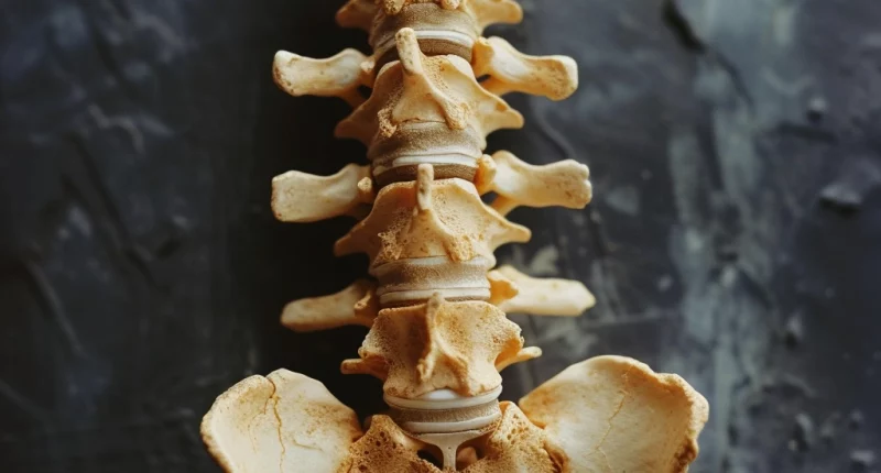
Spondylolisthesis is a displacement of the vertebrae to the front or back due to increased mobility of the spinal column structures. The superior vertebra is displaced.
About the disease
Spondylolisthesis is most commonly found in the lumbar spine, including in the area where the fifth lumbar vertebra joins the first sacral vertebra. Much less often this pathology is found in the cervical region. This topical prevalence of the process is explained by the load experienced by the spinal column (it is maximum in the lumbosacral region).
The main complaints of patients are back pain radiating to the leg. Muscle weakness, rapid fatigability and paresthesias in the lower extremities are accompanying signs. The diagnosis is established on the basis of the data of clinical examination, which is supplemented by imaging diagnostics, primarily radiological. Severe spondylolisthesis is considered a dangerous pathology, as it can lead to severe neurological symptoms.
Treatment of spondylolisthesis is performed by conservative or surgical methods. The choice of treatment program is determined by the degree of curvature of the vertebral column, stability or instability of the displaced vertebra, as well as the severity of neural compression. Surgery is recommended for severe deformities that limit a person’s activity and jeopardize the spinal cord.
Types of spondylolisthesis
Based on radiologic findings, the following degrees of severity of spondylolisthesis are distinguished:
- First degree – the vertebra is displaced by a quarter;
- Second degree – the vertebra is displaced by half;
- Third degree – the vertebra is displaced by three quarters;
- Fourth degree – the vertebra is displaced all the way around;
- The fifth degree is vertebral prolapse, which increases the risk of traumatic injury to the spinal cord.
Symptoms of spondylolisthesis
Symptoms of spondylolisthesis may include the following:
- lower back and sacral pain;
- increased pain sensations with physical activity;
- Irradiation of pain into the legs (this sign is not always positive);
- gait disturbance, limping;
- Intermittent or persistent muscle weakness in the legs;
- a feeling of goosebumps in the lower extremities;
- leg muscle spasm;
- impaired sensation in the feet and lower legs;
- feeling of heaviness in the legs when standing or moving around;
- need to get up at night to walk to relieve uncomfortable symptoms in the legs;
- difficulty lifting the straight leg;
- decrease in skin temperature on one or both legs;
- transient claudication – the need to stop every 10-15 minutes of walking due to the onset of muscle weakness.
Reasons
Classification of types of spondylolisthesis is based on the etiologic pathogenetic mechanisms of disease development. The following varieties are distinguished:
- Spondylolysis (classical, true) type – a defect is first formed in the articular section of the vertebral arch, as a result of which the body of the superior vertebra is displaced to the front (the greater the displacement, the more the defect increases);
- Dysplastic type – slippage of the superior vertebrae develops due to an abnormality of the vertebral column that has existed since birth (unexpanded arch, underdeveloped vertebral body, etc.);
- Degenerative type – dystrophic processes in the intervertebral disc or articular joints lead to displacement of the vertebra;
- Traumatic type – spondylolisthesis can be a direct result of trauma or develop in a delayed period due to secondary deformity;
- Pathological type – slippage of the superior vertebra is caused by primary pathology of the musculoskeletal system (osteogenesis imperfecta, spinal neoplasms, etc.);
- Postoperative type – can occur after spinal surgery when the vertebral arches or arch joints are affected.
Diagnosis
A preliminary diagnosis of spondylolisthesis can be made based on the results of an objective orthopedic examination. Palpation of the spine reveals a depression in the area of a vertebra displaced anteriorly is considered a reliable sign of spondylolisthesis. Other manifestations of this disease may include the following:
- An increase in the anteriorly directed arch in the lumbar spinal column;
- changing the position of the pelvis;
- sacral verticalization;
- flattening of the back;
- curvature of the spinal column in the lateral projection, which is associated with a frequent combination of spondylolisthesis and scoliosis;
- reduced spinal mobility – back-and-forth bending is particularly affected;
- increased tone of the muscles that extend the spine.
Imaging examinations are performed to establish a definitive diagnosis:
- X-ray scan, which is performed in 3 or more planes, including functional tests, including maximal flexion and subsequent maximal extension in the lumbosacral region;
- myelography – X-ray contrast study, which allows you to determine the degree of compression of nerve roots (for contrast injection is performed spinal puncture);
- magnetic resonance scanning – at the present stage is considered the “gold” standard of diagnosis, as it allows you to determine the state of the soft tissue compartment of the spinal column and assess the degree of compression of nerve roots;
- computed tomography – assesses the state of bone structures, so this study is not very informative at the initial stages of the pathological process (unlike magnetic resonance imaging).
Treatment of spondylolisthesis
According to clinical guidelines, treatment of stable spondylolisthetic deformity without complications starts with conservative therapy. Surgical intervention is considered starting from the third degree of spondylolisthesis in the presence of appropriate clinical indications.
Conservative treatment
Conservative treatment has the main goal – to stabilize the spinal-motor segments and control the pain syndrome. It is used in the first or second degree of spondylolisthesis in the absence of pronounced neurological symptoms.
The main thrusts of the conservative program are:
- therapeutic exercise and swimming, which help strengthen the muscular corset that keeps the spine in the correct position;
- electrical stimulation of the muscles involved;
- Taking anti-inflammatory drugs that help to reduce the severity of pain;
- warming procedures, including physiotherapy (has an additional analgesic effect);
- neuroprotective therapy, which reduces the severity of edema of nerve roots and improves their blood supply;
- wearing a corset – recommended during the exacerbation period (stabilization of the spine can reduce the severity of pain, but long-term wear is contraindicated, as it may cause secondary atrophy of paravertebral muscles).
Surgical treatment
Surgery for spondylolisthesis is performed in order to reliably fix the affected spinal segment and subsequent stability. Surgical intervention can be performed from the anterior, dorsal (posterior) or combined. To restore the anatomical structure of the vertebral column, special plates and fixators made of medical titanium are used. A contraindication to surgery is the presence of severe somatic diseases with impaired functional activity of the organ.
All these treatment options are available in more than 820 hospitals worldwide (https://doctor.global/results/diseases/spondylolisthesis). For example, Anterior lumbar interbody fusion (ALIF) is performed in 31 clinics across Turkey for an approximate price of $7.2 K (https://doctor.global/results/asia/turkey/all-cities/all-specializations/procedures/anterior-lumbar-interbody-fusion-alif).
Prevention
Prevention is aimed at timely correction of congenital anomalies of the spinal column. In case of injury, it is recommended to consult a traumatologist as soon as possible to avoid the development of immediate and distant deformities. Proper sitting, strengthening of the muscle corset and regular warm-ups during prolonged sedentary work play an important role in preserving the health of the spine.
Rehabilitation after spondylolisthesis
Postoperative rehabilitation after spondylolisthesis in the early period is aimed at preventing osteoporosis around the installed plate. For this purpose, it is recommended to limit physical activity (but not to adhere to bed rest), as well as to take calcium preparations with vitamin D. As fresh bone tissue appears around the implant, the physical activity regimen is expanded. Therapeutic physical training is carried out under the supervision of a specialist in LFC. For the first month after surgery, prolonged sitting is not recommended. The complex rehabilitation program may also include physical procedures that help to eliminate residual neurological disorders.


