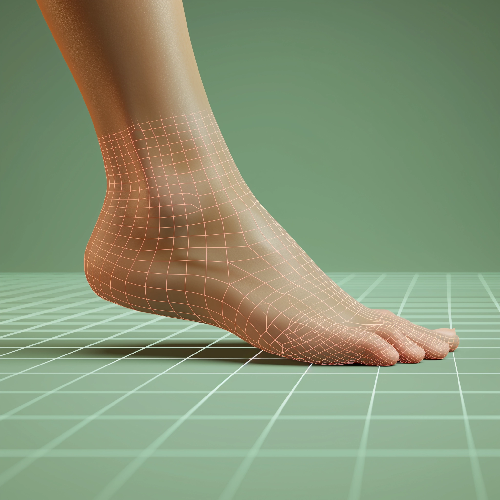

Foot and ankle deformities
What’s that?
Foot deformities are persistent organic changes in the shape and length of the musculoskeletal compartment of the distal segment of the lower extremity.
About the disease
The most common variant of foot deformity is hallux valgus, where the big toe is deviated to the outer edge and, in advanced cases, may overlap the second toe. The deformity is usually a complication of a primary process that results in curvature or shortening of the bony structures of the foot, pathologic changes in the joint, and damage to the tendon-ligamentous compartment.
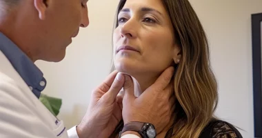
Diagnosis of valgus deformity of the foot is based on assessing the clinical picture, objective examination data, and imaging diagnostic methods. The disease may be accompanied by pain in the foot, which increases when walking and wearing tight shoes. Other manifestations are claudication and pain in the higher joints of the leg and the lower back (due to the increasing load on the spine). Plantography and radiography are performed to assess the bone anomaly present clearly, and a layer-by-layer scan with CT or nuclear magnetic tomography is performed if indicated.
Depending on the stage of the pathological process, treatment may be conservative or surgical. The optimal correction program will be determined by a specialist.
Even though valgus deformity is one of the most common orthopedic pathologies of the foot, other variants are possible (described below in the classification). Therefore, the orthopedic traumatologist’s responsibility is to diagnose and select differentiated treatments for foot deformity.
Types of foot deformities
The following types of foot deformities are distinguished in adults and children:
- static deformations;
- post-amputation deformities;
- Charcot foot (diabetic neuropathic osteoarthropathy) is a specific complication of diabetes mellitus.
The first group includes hammer and claw deformity of the toes, transverse spreading, valgus deviation of the first toe, Taylor’s deformity, limitation of dorsal flexion of the foot, and others.
The second group includes secondary deformities that are due to the need to perform mutilation, such as in patients with diabetic foot or after traumatic tissue crush.
Symptoms
The main clinical signs of deformity may include:
- a visually detectable curvature of the foot;
- pain when walking;
- the difficulty of fitting shoes;
- limitation of mobility in the ankle and mobile joints of the foot;
- rapid development of fatigue when walking and exercising;
- secondary curvature of posture.
By the appearance of the foot, the doctor can make a preliminary diagnosis:
- pronounced flexion toward the sole, in which it is difficult or practically impossible to bend the foot to the opposite side, indicates a horse foot;
- flexion of the distal leg toward the rear (closer to the shin) is a sign of heel foot;
- foot that is so curved that it rests only on the heel tubercle and the heads of the metatarsal bones is called a hollow foot;
- foot that does not have physiologic arches indicates flat feet (in the transverse or longitudinal direction);
- with clubfoot, the foot is shortened and turned outward.
Causes of foot deformities
The following factors can cause foot deformities:
- congenital, including hereditary, anomalies of the musculoskeletal compartment of the distal part of the foot;
- post-traumatic changes that form when a fracture does not heal correctly;
- post-burn changes – acquired foot deformity is caused by third- to fourth-degree burns;
- deforming osteoarthritis of the metatarsophalangeal and interphalangeal joints, which leads to gradual obliteration of the cartilage layer and exposure of bone tissue with the formation of persistent fusions;
- neurological disorders that lead to paresis (partial loss of innervation) or paralysis (complete blockade of neuromuscular conduction) – may be the result of stroke, multiple sclerosis, and other pathologies.
Diagnosis
Diagnosis of the type and degree of foot deformity is based on the results of additional examination, which may include the following methods:
- Plantography – visual or automated determination of the severity of the longitudinal and transverse arch of the foot;
- X-rays – allows you to determine the condition of the bony structures of the foot and ankle;
- Computed tomography or magnetic resonance imaging – performed when it is necessary to obtain layer-by-layer scans of the studied anatomical area.
Treatment of foot deformity
Treatment of foot deformity is determined by its type, cause, and degree of curvature. Conservative measures are recommended for minor deformities, and surgery is indicated for severe deformities.
Conservative treatment
Conservative treatment (shoes, insoles, orthopedic devices) is prescribed based on the need for a particular type of correction and relief.
The footwear to be used by patients with foot deformities is characterized by several features:
- stiff, non-bending sole with a rocker sole or roller sole;
- the width of the shoe is not less than the width of the foot;
- the beveled front edge of the heel (reduces the likelihood of injuries and falls);
- sufficient depth of the model for the possibility of wearing a special insole;
- a specialist should select shoes.
For high-risk patients, it is advisable to use individually made orthopedic shoes and individual insoles, modeling the arch of the foot, more effectively relieving the areas subject to compression.
When indicated, various orthopedic devices (hammertoe correctors, metatarsal cushions, etc.) are used. The selection of orthopedic correctors should only be strictly under the individual control of a specialist. The most common are individually manufactured silicone correctors for finger deformity.
Surgical treatment
Depending on the type of deformity and the severity of clinical signs, the following interventions may be performed:
- resection arthroplasty – allows you to cope with the increased mechanical impact caused by stiffness of the first toe in deforming osteoarthritis of the metatarsophalangeal joint;
- resection of the metatarsal head – used to locally reduce the mechanical impact on the plantar surface in its projection;
- mini-invasive osteotomies of the bones of the foot – a section is made at the level of the distal metaphysis of the metatarsal bone with a special cutter through a puncture in the skin, and the head is displaced to the rear, thus this area is removed from the load;
- tenotomies of the tendons of finger flexors and extensors – performed with a thick needle through skin punctures (this is the least traumatic type of surgical orthopedic treatment);
- Achilles tendon lengthening – aims to reduce pressure on the plantar surface of the entire forefoot.
Reconstructive surgeries for congenital and acquired foot deformities solve two important problems:
- restoration of lost foot function;
- improvement of the patient’s appearance, psychological, and social rehabilitation.
All these treatment options are available in more than 760 hospitals worldwide (https://doctor.global/results/diseases/foot-and-ankle-deformities). For example, complex foot surgery can be performed in 29 clinics across Turkey for an approximate price of $3.9 K (https://doctor.global/results/asia/turkey/all-cities/all-specializations/procedures/complex-foot-surgery).
Prevention of foot deformities
Prevention of foot deformities primarily involves wearing high-quality and comfortable footwear that meets orthopedic requirements (follows the physiological curves of the foot and allows even distribution of the load on the feet). Patients with diabetes mellitus, it is essential to adequately compensate for carbohydrate disorders to reduce the risks of developing diabetic foot, which can cause deformity. Safety precautions and a safe environment for moving children are recommended to prevent burning deformities.
Rehabilitation of foot deformity
In the postoperative period, orthosis support is recommended (wearing a bandage at night for several months), and individual shoes are made and worn. The rehabilitation program for valgus and other foot deformities includes physiotherapy, massage, and physical therapy.