Spinal deformity
What does spinal deformity mean?
Spinal deformity is an abnormal spinal curvature in the frontal or sagittal plane, accompanied by a change in its standard configuration. Treatment of this condition is handled by an orthopedist or spinal surgeon.
About the disease
The prevalence of this disease is attributed to the advent of the so-called “sedentary age.” Today, people study and work sitting down, and it is in the sitting position that the maximum pressure on the spinal column is achieved.
During childhood, the child’s spine undergoes dynamic changes, resulting in physiological curves. These curves arc forward in the cervical and lumbar spine, while in the thoracic and sacral spine, they are directed to the rear. The physiologic nature of the curves is that these curvatures help to reduce the force of vertical loading by transferring it to the paravertebral ligaments. The curvatures help smooth out jolts during multi-intensity motion. The semi-mobile articulation of the vertebrae allows the spine to curve but still maintain the correct shape.
The most common causes of posture deformities occurring in childhood and adolescence are personality traits and heredity, static disorders (constant incorrect postures of the child), and sedentary behavior. Schoolchildren, students, and office workers are forced to spend much time sitting with a slight inclination forward. The consequence of this process is the relaxation of paravertebral muscles and the entire weight of the body taken on the spine. These processes initially lead to poor posture. However, if the impact of unfavorable factors continues, no functional but organic changes occur in the spinal column.
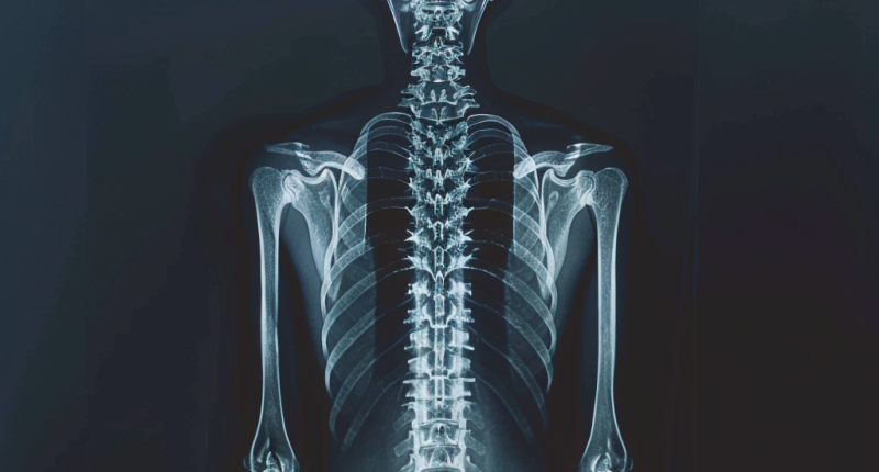
In the initial stage, there are no visible signs of the disease. As the deformity progresses, the appearance of the person changes. Initial diagnosis is based on X-ray scanning data.
Treatment of mild degrees involves a conservative approach. In severe cases, surgical (orthopedic) intervention may be indicated.
Types of spinal deformities
Doctors distinguish three types of spinal deformity:
- Pathological kyphosis – the convex part of the arch is oriented backward (the angle is open to the front);
- pathologic lordosis – the convex part of the arch is directed forward (the angle is open backward);
- Scoliosis is a lateral deformity of the spinal column that can lead to the formation of a rib hump;
- Kyphoscoliosis is a change in the shape of the spinal column in the lateral plane and the front-to-back direction (S-curvature).
A distinction is made between kyphosis and lordosis. If the magnitude of these curves increases, this leads to the formation of pathological deformities. In the thoracic and sacral region, they are oriented backward, and in the cervical and lumbar segments – forward.
In addition to kyphotic and lordotic deformities, kyphosis is distinguished, which is always considered a pathology (the spine in the sagittal plane should not have curvature). Scoliosis is regarded as the most common anomaly, and at the same time, this pathology most often causes deformity in the thoracic region. The direction of scoliosis can be twofold, so the right- or left-sided form is distinguished.
Lateral spinal deformities are categorized into 4 degrees of severity:
- First degree – the angle of spinal deformity varies between 5-10 degrees. In the lateral plane, there is a slight deformity. This stage of the disease is detected only based on X-ray data; it is impossible to notice the change in the shape of the spine with the naked eye.
- The second degree – the angle of vertebral deformation is between 11 and 30 degrees. At this stage, the spine has a significant deviation. The rotation of the vertebrae around its axis leads to the appearance of compensatory arches. When palpating the periorbital area, the doctor can detect a myofascial roll and a rib hump.
- Third degree – the magnitude of the vertebral angle of curvature corresponds to 31-60 degrees. At this stage, there are persistent changes and a more severe change in the shape of the thorax. When conducting X-ray scans, the vertebrae in the deformation zone have the shape of a wedge. The structure of the intervertebral discs is violated.
- Fourth degree – the angle of vertebral deformity exceeds 60 degrees. At this stage, the aesthetics of the body are significantly disturbed, which leads to the formation of a psychological complex in the patient. The bony pelvis is deformed, the body is deviated, the mobility of the spine is disturbed, the shape of the thorax is altered, and a rib hump in front and behind is noted. Radiography visualizes wedge-shaped vertebrae, lesions of intervertebral joints, and deposition of calcium salts in the ligamentous-tendinous apparatus of the spine.
Symptoms of spinal deformities
The nature of the deformity determines symptoms of spinal deformity. For example, in scoliosis:
- is defined as a slouch;
- The shoulder blades, shoulders, and pelvis are at different levels on the left and right sides;
- Visually, one upper limb appears longer than the other;
- When tilting the torso, the rib hump is visualized, the size of the triangles on the waist changes;
- Back pain occurs if a person stays in the same position for a long time or performs heavy physical exercises;
- Rapid fatigue of the paravertebral muscles;
In most cases, scoliosis of the spine will actively progress, and the above symptoms may be added to quite real problems – respiratory disorders, malfunctions of internal organs, and osteochondrosis. People with scoliosis are more prone to bronchial asthma, pneumonia, and problems with the cardiovascular system.
Causes of a spinal deformity
The causes of spinal deformity are classified into two categories – congenital and acquired.
Congenital deformities are formed due to improper laying and development of the spinal discs and ligaments while still in the womb. In early childhood, vertebral deformities can result from rickets, cerebral palsy, and poliomyelitis.
The acquired type of the disease develops during childhood and adolescence, especially during active body growth. Possible causes include:
- weakness of the back muscular corset;
- sedentary;
- unbalanced diet;
- asymmetrical types of physical activity;
- a pronounced forward tilt of the torso when sitting;
- One-way carrying of a bag or backpack.
In adults, spinal deformity can occur due to concomitant musculoskeletal disorders. For example, the risk of scoliosis is increased by deforming hip or knee osteoarthritis.
Diagnosis of a spinal deformity
To determine the existence and severity of spinal deformities, various diagnostic tests can be utilized:
- X-ray (Plain Films): This test utilizes invisible electromagnetic energy beams (X-rays) to generate images of bones. It typically does not capture soft tissue structures such as the spinal cord, spinal nerves, discs, and ligaments and may not reveal certain conditions like tumors, vascular malformations, or cysts. X-rays offer an overall evaluation of bone anatomy, curvature, and alignment of the vertebral column. It helps assess spinal dislocation (spondylolisthesis), kyphosis, and scoliosis, as well as local and overall spine balance. Specific bony irregularities like bone spurs, disc space narrowing, vertebral body fractures, collapse, or erosion can also be detected. Dynamic X-rays (showing spine motion) can be obtained to assess abnormal or excessive movement or instability at affected spine levels.
- Computed Tomography (CT) Scan: A diagnostic imaging procedure combining X-rays and computer technology to create highly detailed images of bones and soft tissues. CT scans provide more comprehensive information than conventional X-rays.
- Magnetic Resonance Imaging (MRI): This technique utilizes a magnet and radio waves to produce detailed images not only of the spine but also of the spinal cord, spinal nerves, and other soft tissues.
Treatment of spinal deformities
Treatment methods for spinal deformities are categorized into conservative and surgical techniques. Physical rehabilitation refers to conservative methods. It can be used as a stand-alone treatment or after surgical methods to recover patients. Its task is to suspend the disease’s progression and stabilize the person’s condition.
Conservative treatment
How do you correct scoliosis and other deformations of the spinal column? The main types of conservative care are:
- health-improving massage;
- therapeutic exercise;
- use of special orthopedic corsets;
- physiotherapeutic procedures;
- taping;
In severe pain, non-steroidal anti-inflammatory drugs may be taken for symptomatic purposes.
Surgical treatment
Reconstructive-plastic operations on the spine are performed in case of pronounced deformities that negatively affect the work of internal organs or lead to a significant violation of the body’s harmony, causing psychological suffering in a person.
Surgical correction is performed in more than 827 hospitals around the world (https://doctor.global/results/diseases/spinal-deformity). The price depends on many factors, including age, health condition, diagnosis, and procedure which is required. For example, surgical correction of scoliosis in adult patients in Israel costs 52,305 USD (https://doctor.global/clinic/soroka-medical-center).
Prevention of spinal deformities
Prevention measures for acquired deformities of the spine are pretty simple; it is important to adhere to them regularly:
- arrange pauses in the process of sedentary work;
- exclude mechanical loads of asymmetric type;
- improve the diet to provide the body with vitamins and minerals;
- engage in preventive physical activity (swimming, skiing, cycling);
- maintain correct posture when doing sedentary work.


