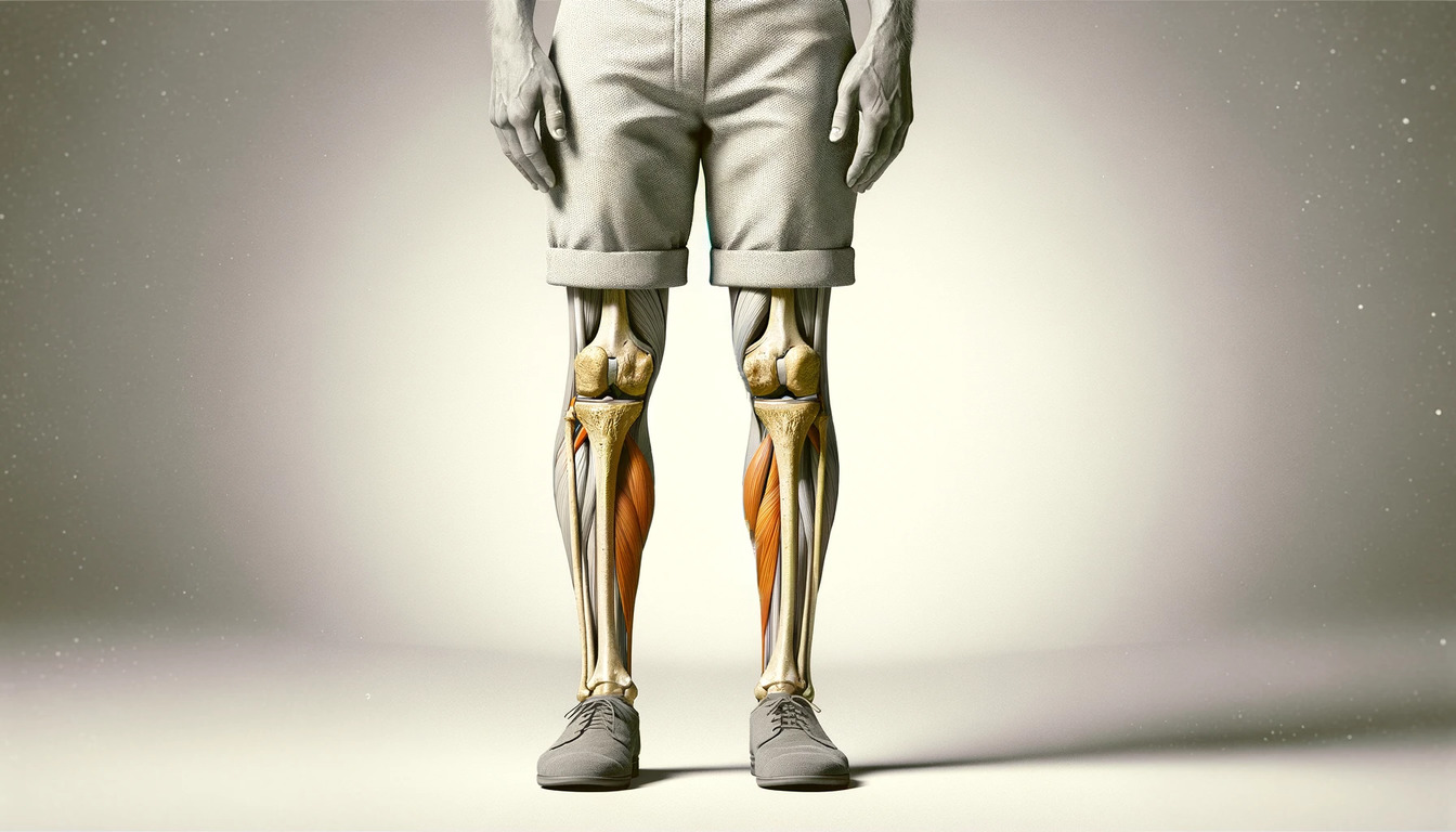

Varus knee
O-shaped legs (varus knee) is a lower limb shape change in which the angle of the knee joint opens outward. Varus knee, commonly known as bowleggedness, is a condition where the knees stay wide apart when a person stands with their feet and ankles together. This condition can affect individuals of all ages, with varying degrees of severity and implications for mobility and joint health. In this comprehensive article, we will explore the causes, symptoms, diagnosis, treatment, and management of varus knee.
About the disease
O-shaped legs are not only a cosmetic disadvantage. In this form of curvature, the anatomical-functional state of the lower limbs may suffer. That happens because the distribution of load on the knee and ankle joints is disturbed. The increased pressure increases the risk of micro-damage with the subsequent development of a degenerative-dystrophic process (osteoarthritis). The O-shaped curvature of the tibia is most often congenital, but in some cases, it can be acquired.
In congenital forms, surgical correction is recommended before the age of 30, as it has a positive effect on the psychological and emotional background of the patient and prevents traumatization of the joints. Different types of surgery can correct Varus deformity of the joints. So, in some cases, immersion plates are used, and in other cases – external fixation devices. However, in both situations, the typical stage is corrective osteotomy – excision of excess bone tissue, which violates the anatomy of the limb.
Types
Varus knee is classified into two categories based on etiology:
- congenital pathology;
- acquired pathology.
Symptoms
Varus deformity of the legs is manifested by rounding the outer contours of the knee – two side-by-side straightened legs resemble the shape of the letter “O.” The degree of curvature may vary.
Other symptoms may include:
- Knee pain or discomfort
- Uneven wear and tear of the shoes
- Difficulty in walking or running
- Stiffness and limited range of motion in severe cases
Causes of varus knee
The causes of varus knee can be as follows:
- congenital traits;
- connective tissue dysplasia;
- vitamin-D resistant rickets – an inherited disease characterized by impaired calcium absorption in the intestine (manifested a few months after birth);
- true rickets (associated with vitamin D deficiency) – risks increase after breastfeeding stops and during adolescence;
- deforming osteoarthritis of the knee;
- Blount’s disease is a lesion of the tibial growth zone;
- Paget’s disease is a violation of the processes of osteogenesis accompanied by bone deformation.
Diagnosis
X-ray scanning objectively diagnoses varus leg deformities in children, men, and women. The method allows you to measure the angles formed by the tibia and femur. Long-axis radiography also helps assess the condition of the femoral neck and hip joint, knee joint, ankle, and foot, including fingers (little finger and others). Laboratory tests are ordered to identify the possible cause of varus deformity. Gait analysis is performed to assess how varus knee affects walking.
Treatment of varus deformity
The specifics of the treatment program are determined by the degree of curvature and the patient’s age. In children, conservative measures are applicable, while surgical interventions with subsequent rehabilitation are preferred in adults.
Conservative treatment
Conservative methods carry out treatment of varus deformity in childhood:
- physical therapy;
- massage;
- wearing special corrective orthotics;
- Use of orthopedic insoles to reduce the load on the foot.
In disorders of phosphorus-calcium metabolism, appropriate drug correction is carried out.
Surgical treatment – surgery for varus deformity
The scope of surgery for varus deformity most often involves a corrective osteotomy (removal of a bone fragment on the larger side) followed by fixation with an external apparatus (Ilizarov apparatus). Corrective osteotomy allows the restoration of standard angles between the femur and tibia. It helps to distribute the load on the joints evenly.
When using external fixation devices, the amount of extension per day can be up to 2 mm. It helps to prevent injury to the ligamentous apparatus of the knee.
Stiffness control is performed after the surgical procedure. After the patient no longer needs dressings daily, the patient can be treated as an outpatient. The external fixation device is removed when clinical and radiological methods confirm the effectiveness. The average period of wearing such a device is 3 to 4 months.
Another option for surgical treatment is corrective osteotomy with the use of immersion plates for bone fixation. In this case, the patient does not wear such a structure as the Ilizarov apparatus in the postoperative period, and the bone segments are fixed with titanium fixators.
All these surgical options are available in more than 900 hospitals worldwide (https://doctor.global/results/diseases/varus-knee). For example, knee osteotomy is performed in 26 clinics across Turkey for an approximate price of $6.0 K(https://doctor.global/results/asia/turkey/all-cities/all-specializations/procedures/knee-osteotomy).
Living with Varus Knee
Managing varus knee involves:
- Regular monitoring, especially in children, to ensure proper leg development.
- Exercises to maintain joint flexibility and muscle strength.
- Wearing appropriate footwear to support the legs and alleviate discomfort.
- In adults with osteoarthritis, weight management can help reduce strain on the knees.
Prevention
Prevention of the disease is aimed at eliminating possible risk factors. While congenital cases cannot be prevented, some measures can reduce the risk of acquired varus knee:
- Ensuring adequate nutrition, especially in children.
- Preventing and promptly treating leg injuries.
- Regular medical check-ups in children for early detection.
Rehabilitation
Patients are prescribed physical procedures, analgesia, and anticoagulant therapy in the postoperative period. These measures aim to prevent deforming arthrosis of the knee and ankle joints. It is also recommended to move the foot to prevent contractures. Activation of patients begins the day after surgery. The mode of activity is expanded gradually. At the first stage, walking with a walker is recommended; at the second stage – with crutches; at the third stage – without additional support.
After the removal of the external fixation apparatus, a course of rehabilitation massage, physical therapy, and physiotherapeutic procedures are carried out. The complex of rehabilitation measures is developed individually for each patient.
Conclusion
Varus knee is a condition with a wide range of causes and varying degrees of severity. While in many cases, especially in children, it resolves on its own, some instances require medical intervention. Understanding the underlying cause is crucial for effective treatment and management. Advances in medical treatments and surgical techniques have significantly improved the outcomes for individuals with varus knee, enabling them to lead active and comfortable lives. It’s important for those affected or caring for someone with varus knee to consult healthcare professionals for proper guidance and treatment strategies.