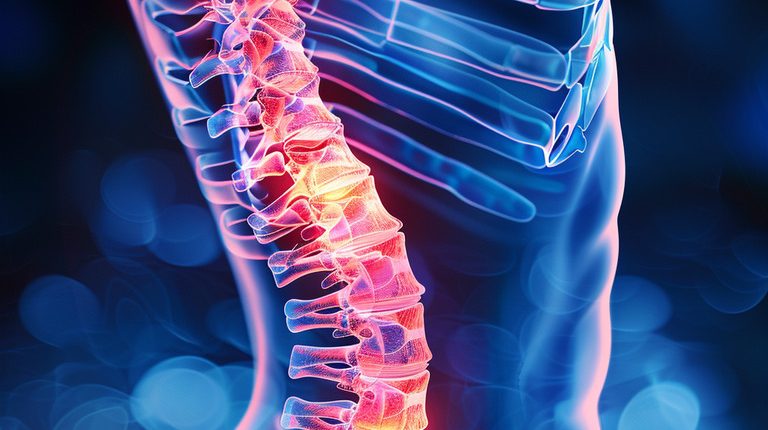
Vertebral compression fracture
What does a spinal compression fracture mean?
A vertebral compression fracture is when one bone fragment is pressed into another bone fragment in the area of the vertebral body.
About the disease
Most compression fractures are secondary in origin, whereby bone strength is reduced, and the vertebral bodies are allowed to push into each other. Rarely, fractures are primary and associated with a high-energy external impact.
What does a spinal compression fracture mean from a diagnostic point of view? It is the identification of a wedge-shaped defect in the structure of the vertebral body. Clinically, such a condition is accompanied by pain in most cases. However, there can sometimes be asymptomatic forms that are discovered incidentally when radiographs are taken on another occasion. Imaging methods also make it possible to confirm that the fracture has become consolidated against the background of ongoing treatment.
Treatment can be carried out by conservative and surgical methods. The first option is symptomatic, and the second – in addition to controlling pain, also helps to prevent the progression of the pathological process and reduce the risk of complications.
Types of spinal compression fractures
According to localization, the following types of compression fracture of the spine are distinguished:
- disruption of the integrity of the cervical vertebrae;
- disruption of the integrity of the thoracic vertebrae;
- disruption of the integrity of the lumbar vertebrae;
- disruption of the integrity of the sacral vertebrae.
Most often, vertebral compression fractures occur where the vertebral column has normal curves designed to reduce mechanical loading. The seventh and eighth thoracic vertebrae and the twelfth thoracic and first lumbar vertebrae are considered the most “dangerous” places.
Three degrees of depressed fractures are distinguished based on the reduction in vertebral body height:
- first, the vertebral body sags 30%;
- second – the value of drawdown from 30 to 50%;
- third – the value of the drawdown is more than 50%.
Symptoms of a spinal compression fracture
The nature of their origin determines symptoms of spinal compression fracture. In the case of a pathological fracture, clinical manifestations develop gradually. Pain sensations at the beginning are insignificant, but gradually, their intensity increases and becomes constant. When the nerve roots passing in the intervertebral foramen are compressed, paresthesiasand numbness of the upper or lower extremities may appear (depending on the compression level). Osteoporotic vertebral degeneration fractures may be characterized by stooping or humping.
Traumatic fractures have vivid symptomatology and a clear indication of the impact of a mechanical factor. If a person has a sudden pain, breathing may be disturbed (when thoracic vertebrae are damaged). In the horizontal position of the body, pain decreases and increases during coughing or when trying to move.
Causes of spinal compression fracture
The most common cause of spinal compression fracture in elderly patients is osteoporosis (decreased bone mineral strength). Other causative factors may include:
- Hemangiomas are vascular tumors that exert mechanical pressure on bone tissue and thus reduce its strength;
- Spinal disc hernia is a pathologic condition in which the intervertebral disc bulges toward the vertebral body;
- Metastatic lesion of the vertebrae by malignant tumors.
In some cases, compression fractures are associated with exposure to a high-energy traumatic factor. This mechanism usually occurs in young patients in automobile accidents or falls from heights.
Diagnosis of spinal compression fracture
Diagnosis of a compression fracture of the spine is based on imaging examination methods:
- X-ray scan;
- magnetic resonance imaging (MRI).
These tests help to determine the height of the vertebral bodies. In depressed fractures, it is reduced by 1/5 or more either throughout or in a specific location. This is what a compression fracture looks like, i.e., there is no typical line of bone damage but usually a wedge-shaped defect.
In some cases, it is necessary to distinguish an old fracture from a fresh one. A CT scan can help answer this question.
In the presence of fractures on the background of hemangiomas, it is recommended to clarify the prevalence of pathologic lesions and perform prophylactic treatment of active foci. If the metastatic nature of a vertebral fracture is suspected, scintigraphy is indicated, which helps to identify the main focus of the lesion.
Treatment of spinal compression fracture
The choice of treatment method for a spinal compression fracture is determined by the degree of damage to the vertebral body, the integrity of the closure plate (a kind of bone and cartilage enveloping the vertebral body), and the presence of splinters.
Conservative treatment
Conservative treatment of vertebral fractures in adults is aimed at controlling pain and improving the quality of life. Such therapy consists of the following components:
- medication analgesia – systemic (injectable or oral NSAIDs) or local (blockages);
- immobilization of the pathologically altered segment with orthopedic orthoses;
- physical therapy (if there are no contraindications);
- calcium preparations and bisphosphonates, which help to increase the mineral strength of bone tissue.
Surgical treatment
The indications for surgery for a spinal compression fracture are as follows:
- progressive decrease in vertebral body height;
- curvature of the spinal column;
These indications are relevant even when conservative therapy helps to reduce the pain syndrome. Surgical intervention will prevent possible adverse effects and will have a positive impact on the quality of life.
- Ineffectiveness of conservative treatment for pain relief.
The development of side effects in response to long-term anesthetic therapy is also a relative indication for surgery.
Current methods of surgical treatment of compression uncomplicated fractures are:
- vertebroplasty;
- balloon kyphoplasty.
These methods can reduce or eliminate pain syndrome and stabilize the spinal column. Such interventions are minimally traumatic and well tolerated by patients. Currently, the indications for vertebroplasty or balloon repair have been expanded – in some cases, the surgery is performed to prevent the occurrence of a spinal fracture.
- Vertebroplasty allows you to strengthen the vertebra at the fracture site by injecting medical cement through a special cannula.
- Balloon kyphoplasty is characterized by inserting a catheter into the body of a broken vertebra, which is then expanded. It increases the vertebra’s height, and subsequently, bone cement is injected into the cavity formed. Thus, this method helps to correct the existing deformity of the vertebral bodies.
If the fracture is unstable, open fixation of the fragments is performed to prevent complications. Special metal structures are used for this purpose. In cases where the vertebra is almost wholly destroyed, bone grafts are used.
The period of fracture treatment and subsequent rehabilitation is individualized for each patient. The initial stage of recovery is usually at least one month.
All these treatment options are available in more than 600 hospitals worldwide (https://doctor.global/results/diseases/vertebral-compression-fracture). For example, Vertebroplasty can be performed in 18 clinics across Turkey for an approximate price of $4.2 K (https://doctor.global/results/asia/turkey/all-cities/all-specializations/procedures/vertebroplasty).
Prevention
According to clinical guidelines, preventive determination of bone mineral strength by densitometry is recommended to prevent osteoporotic fractures of the vertebral column. The risk of fractures increases if conditions such as osteopenia and osteoporosis are detected.
Rehabilitation after spinal compression fracture
Rehabilitation after a spinal compression fracture is aimed at improving the functionality and mental and bodily health of the patient. The rehabilitation program soon after surgery may include the following areas:
- providing functional rest;
- wearing orthotics;
- analgesia with medication and non-medication methods.
At the same time, the length of stay in bed after surgery should be as short as possible. That is because muscle hypotrophy begins to develop after four days of being in a horizontal position.
A few days after surgery, a gradual expansion of the rehabilitation program is carried out:
- breathing exercises and performing physical exercises that do not require special exertion;
- physical therapy and acupuncture;
Recently, methods of apparatus mechanotherapy with biofeedback effect have been actively introduced into rehabilitation practice. This method involves exercising on high-tech simulators specially designed to consider the musculoskeletal system’s biomechanics.


