Definition and etiology
Arthrofibrosis is a painful growth of connective tissue inside the ankle joint. These growths can also limit the mobility of the ankle.
Scarring and arthrofibrosis can be caused by surgery, trauma (prolonged immobilization), or joint infections. Adhesions can also form due to damage to the lateral ligamentous apparatus.
There are cases when extensive intra-articular adhesions and fibrosis of the anterior capsule form after open surgical interventions on the ankle joint (after open repositioning and internal fixation for fractures of the tibia, talus, or lateral ankle).
Clinical picture
The symptoms of ankle arthrofibrosis are pretty varied. The most common are:
- Pain at the beginning of movements
- Morning pain at rest
- Rapid fatigue and weakness during physical exertion
- Reducing walking distance
- Friction noise in the joint
- Impaired motor function (impingement syndrome)
- Increased stiffness in the joint
- Swelling and inflammation
- Joint deformity
- Joint locking due to soft tissue, articular cartilage, and bone particles being trapped in the joint.
Diagnosis
The volume of movement in the joint is assessed, and areas of pain are palpated. No clinical tests are performed to determine the exact location and extent of scarring.
An intra-articular injection of local anesthetic may help diagnose the condition. If only a few milliliters of solution can be injected into the joint, this indicates extensive fibrosis that will make arthroscopic and instrumented ports difficult to establish.
The consequences of trauma or surgery may be detected. However, it is impossible to determine the extent of adhesions from radiographic data.
MRI may be helpful in assessing the severity of fibrosis and thickening of the anterior capsule and detecting anterior joint narrowing.
Arthroscopic findings range from isolated scar tissue crossing the joint cavity to almost complete obliteration of the joint cavity by scar tissue. It should be noted that the prevalence of scarring does not always determine the extent of movement restriction. Sometimes, the presence of reasonably extensive intra-articular adhesions leads to only a slight restriction of movement.
Treatment of arthrofibrosis
Conservative treatment
First of all, specialists try to carry out treatment without surgery. The treatment that will be offered to the patient depends on the stage of osteoarthritis, and its goal is to stop or suspend the course of the disease. The most effective methods of conservative therapy:
- Orthopedic shoes
- Chondroprotective treatment to preserve joint cartilage.
- A special orthosis designed to relieve damaged joints in varus or valgus deformity of the foot.
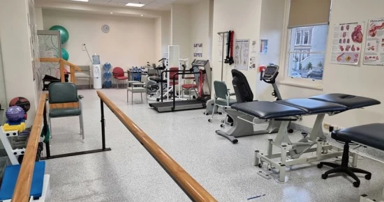
- A set of muscle recovery exercises.
- Acupuncture
- Therapeutic gymnastics
- Physiotherapy
Conservative treatment (treatment without surgery) aims to slow down the course of the disease. Such treatment is mainly a means of restoring joint stability. Physiotherapeutic treatment of osteoarthritis, especially in a specialized orthopedic or traumatology office, with a unique set of exercises, helps to improve the ankle’s condition. In addition, physiotherapeutic treatment accelerates the process of cartilage regeneration after cartilage damage.
Another effective method is to wear special orthopedic shoes that reduce pain, compensate for walking disorders, and optimize joint positions.
Depending on the diagnosis, specialists apply various surgical treatments to slow the disease process or repair existing damage.
It is also treated with anti-inflammatory drugs, so-called anti-rheumatic drugs, or non-steroidal anti-rheumatic drugs (NSAR).
Doctors suggest surgical treatment if conservative treatment has not brought the desired results.
Persistent pain and limitation of motion in patients with a relatively normal radiologic picture may be caused not only by impingement syndrome but also by cartilage damage.
In a significant proportion of these patients, extensive intra-articular scarring causes severe clinical symptoms by creating tension in the anterior capsule. Fibrosis of the anterior capsule of the ankle joint is often a consequence of trauma or surgery.
In this case, the volume of the anterior regions of the joint is reduced, which leads to soft tissue impingement between the talus and tibia. In addition, scars can cause tension in the “sensitive” joint capsule.
Surgical treatment
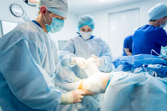
In cases of severe fibrosis, arthroscopic resection of scar tissue with release of the anterior capsule of the joint is the preferred treatment method.
Associated injuries (deep cartilage damage, bone deformities) have an unfavorable effect on the prognosis. Nevertheless, it is surprising how much joint mobility improves in individual cases after arthroscopic arthrolysis, even with severe cartilage damage.
Postoperative management
Patients with localized scarring often report reduced pain immediately after surgery, and joint motion is more accessible. A partial load of 50% of body weight is allowed for 2-4 days, followed by a gradual transition to a full load. Intensive but gentle rehabilitation therapy is recommended to stretch the joint capsule. Vigorous exercise is contraindicated as it may lead to a new fibrotic reaction in the anterior capsule.
As with the postoperative management of patients with arthrofibrosis in the knee joint, close cooperation between surgeon and physiotherapist is necessary. Therefore, inpatient treatment in a particular rehabilitation center is recommended. Jumping and weight-bearing exercises are prohibited for the first 4-6 weeks to avoid unwanted irritation of the capsule.
Rehabilitation
After ligament damage, 20% of patients experience chronic instability in the ankle. Above all, this condition can be detrimental to athletes, as the propensity for injury increases in this case. Ligament instability causes the risk of re-stretching due to injury. Low congruence of joint surfaces can also lead to ankle degeneration, which is a cause of injury and arthritis. Special training to maintain protective reflexes can help prevent further sprains. Please note that special exercises to stabilize the ankle joints should be performed even after surgical reconstruction of the ligaments.
Orthopedic shoes and orthopedic insoles create the conditions for correct loading. Deformities can be corrected by increasing the height of the outer or inner edge of the shoe. In this way, ergonomic footwear helps to reduce joint pain and halts the progression of the disease.
In addition, a special ankle orthosis is used to fix the foot in a given position (correction of internal or external rotation). Orthotics help reduce pain and improve mobility at the initial stage of the disease.

