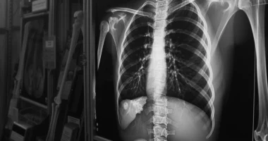
Hip dysplasia
Definition
Hip dysplasia is a congenital disorder of the joint formation process that can cause dislocation or subluxation of the femoral head. There is either underdevelopment of the joint or increased mobility in combination with connective tissue deficiencies. At an early age, it manifests in the asymmetry of skin folds, shortening, and hip extension limitation. Subsequently, there may be pain, lameness, and increased limb fatigue. Pathology is diagnosed based on characteristic signs, ultrasound data and radiological examination. Treatment is carried out using special fixation devices and exercises for muscle development.
General information
Hip dysplasia (from Greek dys – disorder, plaseo – forming) is a congenital pathology that can cause hip subluxation or dislocation. The degree of joint underdevelopment can vary enormously – from gross abnormalities to increased mobility combined with weakness of the ligamentous apparatus. Hip dysplasia must be detected and treated early – in the first months and years of a baby’s life to prevent possible negative consequences.
Hip dysplasia is one of the widespread congenital pathologies. According to experts in traumatology and orthopedics, the average incidence is 2-3% per thousand newborns. There is a dependence on race: African Americans have it less often than Europeans, and American Indians have it more often than other races. Girls have the disease more often than boys (about 80% of all cases).
Causes
Several factors cause dysplasia. There is a clear hereditary predisposition—this pathology is ten times more likely to be observed in patients whose parents suffered from a congenital disorder of the development of the hip joint. The probability of developing dysplasia is ten times more likely with a breech presentation of the fetus. In addition, the likelihood of this pathology increases with toxicosis, medication correction of pregnancy, large fetus, low water supply, and some gynecological diseases in the mother.
Researchers have also noted a link between the incidence of the disease and unfavorable environmental conditions. Dysplasia is observed 5-6 times more often in ecologically unfavorable regions. The development of dysplasia also affects the national traditions of swaddling babies. In countries where newborns are not swaddled, and the child’s legs are in the position of abduction and flexion for a significant part of the time, dysplasia is less common than in countries with a tradition of tight swaddling.
Symptoms of dysplasia
Hip dysplasia is suspected when there is shortening of the hip, asymmetry of the skin folds, and limitation of hip extension. Asymmetry of the inguinal, hamstring, and gluteal skin folds is usually better detected in children older than 2-3 months. Differences in the folds’ level, shape, and depth are noted during the examination.
The symptom of hip shortening is more reliable in diagnostic terms. The child is placed on the back with legs bent at the hip and knee joints. The location of one knee is lower than the other, indicating the most severe form of dysplasia – congenital hip dislocation.
Another symptom indicative of joint pathology is restricted movement. In healthy newborns, the legs are withdrawn to a position of 80-90° and rest freely on the horizontal surface of the table. If the extension is limited to 50-60°, there is reason to suspect congenital pathology. In a healthy baby of 7-8 months of age, each leg is withdrawn to 60-70°, and in a baby with congenital dislocation, to 40-50°.
Complications
If the changes are minor and untreated, there may be no painful symptoms at a young age. Later, at 25-55 years, dysplastic coxarthrosis (hip joint arthrosis) may develop. As a rule, the first symptoms of the disease appear against the background of reduced motor activity or hormonal changes during pregnancy.
Characteristic features of dysplastic coxarthrosis are acute onset and rapid progression. The disease is manifested by unpleasant sensations, pain, and restriction of movement in the joint. Movement in the joint is sharply limited. In the initial period of the disease, the greatest effect is provided by adequately selected physical activity. With a pronounced pain syndrome and malposition of the hip, endoprosthesis is performed.
In unrepaired congenital hip dislocation, a new incomplete joint is formed over time, combined with limb shortening and muscle dysfunction. This pathology is currently rare.
Diagnosis
A preliminary diagnosis of hip dysplasia can be made as early as in the maternity hospital. In this case, it is necessary to visit a pediatric orthopedist within three weeks, who will conduct the necessary examination and make a treatment plan. In addition, to exclude this pathology, all children are examined at 1, 3, 6, and 12 months.
Special attention is paid to children at risk. This group includes all patients with a history of maternal toxemia during pregnancy, large fetuses, and breech presentation, as well as those whose parents also suffer from dysplasia. If signs of pathology are detected, the child is referred for additional tests.
Clinical examination of the baby is carried out after feeding in a warm room in a calm, quiet environment. To clarify the diagnosis, such techniques as radiography and ultrasonography are used. In young children, a significant part of the joint is formed by cartilage, which is not displayed on radiographs, so until the age of 2-3 months, this method is not used, and later, in reading the images, special schemes are used. Ultrasound diagnostics is an excellent alternative to radiologic examination in children in the first months of life. This technique is safe and informative.
It should be remembered that the results of additional tests alone are not sufficient to diagnose hip dysplasia. The diagnosis is made only when both clinical signs and characteristic changes on radiographs and/or ultrasonography are detected.
Treatment of hip dysplasia
Treatment should begin as early as possible. Various devices are used to hold the child’s legs in flexion and extension: splints, stirrups, pants, and special cushions. Only soft elastic structures that do not interfere with limb movements are used in treating children in the first months of life. Wide swaddling is used when complete treatment is not possible, as well as in the treatment of babies at risk and patients with signs of immature joints detected by ultrasonography.
Special muscle strengthening exercises are essential in restoring range of motion and stabilizing the hip joint. A separate set of exercises is prepared for each stage (leg extension, joint retention, and rehabilitation). In addition, massage of the gluteal muscles is prescribed for the child during treatment.
In severe cases, a one-stage closed repositioning of the dislocation is performed with subsequent immobilization with a plaster cast. This manipulation is performed in children ages 2 to 5-6. When the child ages 5-6, repositioning becomes impossible. In some cases of high dislocations in patients aged 1.5-8 years, skeletal traction is used. If conservative therapy is ineffective, corrective surgeries are performed: open reduction of the dislocation and surgical interventions on the acetabulum and the upper part of the femur.
All these treatment options are available in more than 770 hospitals worldwide (https://doctor.global/results/diseases/hip-dysplasia). For example, Hip osteotomy can be performed in these countries for following approximate prices:
Turkey $4.8 K in 14 clinics
China $17.5 K in 6 clinics
Germany $18.2 K in 35 clinics
Israel $24.2 K in 13 clinics
United States $28.4 K in 15 clinics.
Prognosis and prevention
The prognosis is favorable with early treatment and timely elimination of pathological changes. In the absence of treatment or with insufficient effectiveness of therapy, the outcome depends on the degree of hip dysplasia. There is a high probability of early development of severe deforming arthrosis. Prevention includes examination of all young children and timely treatment of detected pathology.

