
Sternal fracture
A fracture of the sternum, or breastbone, situated in the middle of the chest, is known as a sternal fracture. This type of injury happens in about 5-8% of individuals who suffer severe blunt trauma to the chest. Sternal fractures often occur during vehicular accidents when the chest collides with the steering wheel or dashboard or when a seatbelt impacts it. These fractures are categorized as a form of closed chest trauma and frequently occur alongside fractures of the ribs or clavicle. The sternum is a flat, spongy bone to which the rib cartilages are connected at the front of the chest.
Causes
Sternal fractures primarily result from blunt trauma to the chest and deceleration injuries, with common causes including motor vehicle accidents, sports injuries, falls, and physical assaults. These incidents often lead to anterior chest wall pain.
Key points to consider:
- Blunt chest trauma is the primary factor behind sternal fractures.
- Cardiopulmonary resuscitation, sports-related injuries, falls, and assaults are significant contributors to traumatic cases.
- Individuals with significant thoracic kyphosis, osteoporosis, or osteopenia may develop insufficiency fractures of the sternum.
- Certain groups at higher risk include those on long-term steroid therapy, postmenopausal women, and older people.
- Stress fractures of the sternum, though less common, can occur due to repetitive strain, particularly in athletes or military personnel.
Anatomy of sternum
The sternum, also known as the breastbone, is a flat, elongated bone at the front of the chest. It is a crucial component of the ribcage and plays a vital role in protecting the underlying organs, such as the heart and major blood vessels. The sternum consists of three main parts:
- Manubrium: The uppermost portion of the sternum, the manubrium, is shaped like a broad, triangular plate. It articulates with the clavicles (collarbones) and the first two pairs of ribs.
- Body (or Gladiolus): The body of the sternum is the middle section and is longer and narrower than the manubrium. It connects to the third to seventh pairs of ribs.
- Xiphoid Process: The xiphoid process is the sternum’s smallest and most inferior part. It is a thin, cartilaginous extension at the lower end of the sternum. The xiphoid process fully ossifies (turns into bone) in adulthood.
The upper section has a sternoclavicular joint, which articulates the clavicle and the manubrium. Bone-cartilaginous connections of the ribs with the sternum are pretty plastic because their primary functional purpose is to provide unimpeded excursions of the thorax. This anatomical structure determines the rarity of sternal fractures, which account for no more than 5% (of the total structure of fractures). However, if fractures do occur, they usually involve the area where the manubrium joins the body. They are usually caused by a strong compressive force applied externally.
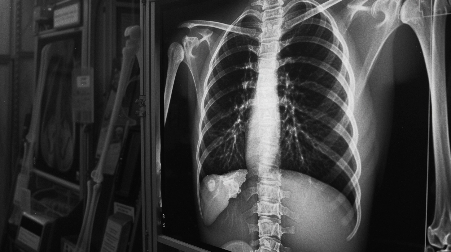
Diagnosis
The final diagnosis of a sternal fracture is based on radiologic examination. X-ray scans are performed in at least two projections. The state of the lung tissue is assessed since the pleural fields are well visualized. In the lateral projection, the condition of the sternum is determined, and the line of bone damage is detailed. In complex clinical cases, computed tomography (CT) may be indicated to detect hidden signs.
As part of a comprehensive examination of patients with sternal fractures, other methods are also performed:
- electrocardiography – assesses the state of cardiac activity;
- ultrasound scanning of the heart with Doppler ultrasonography.
Symptoms
Symptoms of a sternum fracture may include the following:
- intense pain in the area of the damaged bones;
- increased pain when breathing and feeling the chest;
- swelling of soft tissues;
- hemorrhages in the soft tissues;
- localized change in the correct shape of the chest.
Treatment
Both conservative and surgical methods can be used to treat a sternum fracture.
Conservative treatment
With an uncomplicated fracture, fixation of bone fragments is carried out with a wide-band leukoplasty; it is recommended to wear a special rigid corset.
Surgical treatment
Surgical treatment of sternal fracture is indicated in the cases of complete transverse displacement of the fragments.
The traumatologist performs a linear incision and provides access to the injured area. Small bone fragments are removed, and the hematoma is emptied. In the presence of displacement, open repositioning of the fragments is performed with subsequent fixation with plates and screws. In some cases, intraosseous metal osteosynthesis with nailing or plates is performed.
Treatment for sternal fractures is available in over 835 orthopedic clinics globally (https://doctor.global/results/diseases/sternal-fracture). For instance, the cost of surgically treating sternal fractures in Kazakhstan is approximately 1838 USD (https://doctor.global/results/asia/turkey/all-cities/all-specializations/procedures/brain-cyst-surgical-treatment).
Rehabilitation
When the fracture becomes consolidated, it is recommended to perform breathing exercises. Physiotherapy is also helpful.
Complications
Complications like pneumothorax or hemothorax can arise when there is a concurrent injury to both the sternum and ribs. Pneumothorax involves air accumulating in the pleural cavity, while hemothorax is characterized by the presence of blood in the same area. These conditions are severe because they compress the lung tissue, interfering with the lungs’ ability to function properly in ventilation. Immediate medical intervention is essential in these situations.
Prevention
Prevention of sternum fractures is associated with compliance with safety measures – wearing belts during automobile driving and careful work at traumatic production (metallurgical, machine-building).