Aseptic necrosis
Definition
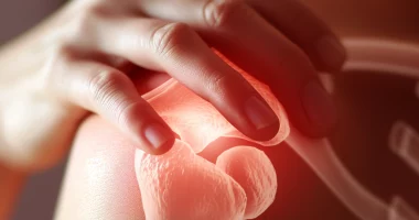
Aseptic necrosis is a severe, chronic disease that destroys bone tissue and limits joint mobility over time. The term “aseptic necrosis” refers to the destruction and death of bone tissue in the absence of infection by pathogens.
Of all bone and muscle diseases, aseptic femoral head necrosis accounts for 1.2 to 4.7%. According to different authors, the disease mainly affects men aged 30-50 years (about 7-8 times more often than women).
In 50-60% of cases, both legs are affected; the process that started on one side occurs on the other in 90% of cases a year later.
Causes of aseptic necrosis:
Aseptic necrosis occurs from the following causes:
✓ hip dislocations, fractures of the femoral neck and the femoral head itself, and surgical interventions;
✓ prolonged use of corticosteroids, especially those intended for systemic use (tablets);
✓ alcohol abuse, leading to metabolic disorders;
✓ prolonged use of painkillers and NSAIDs;
✓ autoimmune diseases, including scleroderma, hemorrhagic vasculitis, and systemic lupus erythematosus;
✓ caisson disease, which is common in divers, miners, and other occupations that require working in rapidly depressurized inhaled gas mixtures;
✓ radiation sickness or radiation therapy to suppress a malignant tumor;
✓ cauda equina syndrome due to impingement of spinal roots at the level of the lumbosacral vertebrae.
Stages of aseptic necrosis.
Symptoms of aseptic necrosis. The primary symptom that occurs in all patients is pain in the affected area. It can vary in intensity depending on the stage of the pathological process.
Stage 1 (pre-radiologic): There are no radiologic changes. It may be asymptomatic or accompanied by pain, muscle atrophy, and restricted movement.
Stage 2 (impression fractures) – the radiograph shows homogeneous darkening, absence of structural pattern in the affected area, local thickening and reduction in the height of the head, and widening of the articular gap. A large number of microfractures are detected.
Stage 3 (sequestration): The head becomes even flatter and loses its normal contours, and the articular gap continues to widen. The images visualize individual bone fragments of different sizes and shapes without normal structure. Thickening and shortening of the femoral neck are detected.
Stage 4 (repair): Bone fragmentation disappears. The femoral head is normally contoured, but its structure must be restored.
Stage 5 (deforming osteoarthritis) – the bone structure is restored, but the head deformity in stages 2 and 3 remains. The head is flat, enlarged, and does not match the shape of the joint socket.
Symptoms of aseptic necrosis.
The primary symptom that occurs in all patients is pain in the affected area. It may vary in intensity depending on the stage of the pathological process.
✓ pain syndrome;
✓ claudication;
✓ visual reduction of muscles in the area of the affected joint;
✓ restriction of free movement in the affected hip joint.
Varieties of aseptic necrosis
Aseptic necrosis of the femoral head
The head of the femur is considered a problem area, where there is an increased risk of arterial blockage, accumulative damage caused by overload, and household trauma. The presence of various pathological processes can provoke aseptic necrosis of the femur.
The leading causes can be attributed to a prolonged intake of hormonal drugs and antibacterial agents. A provoking factor can be the presence of congenital dislocation of the hip, osteopenia, osteoporosis, systemic lupus erythematosus, Bechterew’s disease, and rheumatoid arthritis.
Aseptic necrosis of the knee joint
Destructive processes in the knee joint appear as a result of trauma and loss of blood supply. Joints lose their functions; a person can become disabled. With aseptic necrosis of the knee joint, a person experiences severe painful sensation and observes that the motor ability of the knee is reduced. With the help of magnetic resonance imaging and bone scanning, the initial stage of changes can be detected, as a result of which further loss of bone mass is prevented. This type of treatment is performed in 79 clinics all around the world (https://doctor.global/results/diseases/aseptic-necrosis-of-the-patella).
Aseptic necrosis of the talus.
Necrosis of the talus occurs spontaneously and progresses rapidly. With degenerative changes in the ankle joint, deforming arthrosis develops. In such a situation, mosaic osteochondroplasty is applied, and the joint’s anatomy is restored. You can find all the hospitals, that treat this condition here:
https://doctor.global/results/diseases/aseptic-necrosis-of-the-ankle
Aseptic necrosis of the humerus
This condition causes painful sensations in the shoulder joint, limits its movement, and increases the risk of atrophy. A rare phenomenon is the altered structure of the humerus. If the disease progresses, the patient is prescribed a surgical intervention called endoprosthesis. In this situation, it is the only way to restore the lost function of the upper limbs.
This condition can be treated in more than 790 hospitals worldwide (https://doctor.global/results/diseases/aseptic-necrosis-of-the-shoulder).
Aseptic necrosis of the elbow
Aseptic necrosis of the elbow, also known as osteonecrosis, refers to the death of bone tissue in the elbow joint due to a lack of blood supply. This condition can lead to pain, stiffness, and loss of joint function. It often affects people with certain medical conditions, such as blood disorders, trauma, or long-term steroid use, which can disrupt the blood flow to the bone. Early diagnosis and treatment are crucial to prevent joint damage and preserve elbow function. You can find more information regarding the treatment of this condition here:
https://doctor.global/results/diseases/aseptic-necrosis-of-the-elbow
Aseptic necrosis of the wrist
Aseptic necrosis of the wrist involves the death of bone tissue in the wrist due to insufficient blood supply. This condition can cause significant pain, reduced mobility, and eventual joint collapse if left untreated. Commonly affecting the lunate bone in the wrist, it may result from trauma, steroid use, heavy alcohol consumption, or certain medical conditions that impair blood flow. Early detection and management are vital to prevent progression and maintain wrist function. It is a quiet rare condition, nevertheless, you can find hospitals, that can help with this disease here: https://doctor.global/results/diseases/aseptic-necrosis-of-the-wrist
Diagnosis of aseptic necrosis
Diagnosis of aseptic necrosis is based on a thorough analysis of the patient’s complaints and history of the disease. During the consultation, the orthopedist examines the affected joint and assesses its condition and the degree of dysfunction.
✓ radiography of the affected joint;
✓ a computed tomography (CT) scan;
✓ magnetic resonance imaging (MRI).
Treatment
In the initial stages of the pathological process, drug treatment is performed. At later stages, endoprosthesis may be indicated.
Conservative treatment is aimed at slowing down the processes of bone destruction and improving the patient’s quality of life. For this purpose, the orthopedist applies the following set of remedies:
-anesthetic drugs means that improve microcirculation and nutrient delivery to the damaged area of the bone;
-physiotherapy;
-therapeutic gymnastics.
Operative treatment
At a certain point, the conservative approach becomes ineffective due to the pronounced destruction of bone tissue. In this case, the only treatment option is endoprosthetic surgery of the affected joint. This surgery involves installing an artificial femoral head to completely replace the destroyed one, making it possible to almost completely restore the joint’s function.
All these treatment options are available in more than 60 hospitals worldwide (https://doctor.global/results/diseases/aseptic-necrosis). For example, revision can be performed in 7 clinics across India for an approximate price of $6,1K (https://doctor.global/results/asia/india/all-cities/all-specializations/procedures/revision).
Rehabilitation
Surgical treatment of aseptic necrosis involves replacing the joint with an endoprosthesis. After surgery, the patient remains in the hospital for 1-2 weeks under the constant supervision of orthopedists. The operated limb is immobilized. Every day, the doctor performs dressings and monitors the patient’s condition. To prevent bacterial complications, the patient is prescribed a prophylactic course of antibiotic therapy. To eliminate discomfort, the patient takes painkillers.
Before discharge, the orthopedist teaches the patient how to behave appropriately in everyday life. An individualized plan of rehabilitation measures is developed to restore joint function as quickly as possible. Sutures are removed on the 14th-21st day after surgery. Complete recovery may take up to 3-6 months. It would help if you avoided physical overstrain, ate a complete diet, and rested more during this time.