Becker muscular dystrophy
Definition
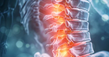
Becker muscular dystrophy is a variant of hereditary X-linked muscular dystrophy characterized by a more delayed and benign course. The disease is characterized by progressively worsening and spreading muscle weakness, hypotonia, and atrophy, initially occurring in the thighs and pelvic girdle muscles. Diagnostic search includes neurologic examination, consultation with a geneticist and cardiologist, neurophysiologic testing of the neuromuscular apparatus, DNA diagnosis, muscle biopsy with morphologic, immunologic, and histochemical study of the obtained samples. Treatment is symptomatic and, unfortunately, ineffective. Progression of the disease leads to the loss of patients’ ability to move independently by the age of 40 years.
General information
Progressive Becker muscular dystrophy was first described in 1955 as a benign variant of Duchenne muscular dystrophy. Subsequently, numerous studies in clinical neurology, genetics, and biochemistry have revealed significant differences in these diseases’ course, biochemical, and morphologic basis. As a result, Becker’s clinical form was distinguished as an independent nosology.
Becker muscular dystrophy is a member of the group of myopathies (myodystrophies), diseases arising from disorders of the structure and metabolism of muscle tissue and manifested by muscle weakness. The pathology is inherited recessively X-linked, so only males are affected. The incidence is one newborn per 20,000 children.
Reasons
The disease is based on a mutation in the gene responsible for encoding the protein dystrophin. About 30% of the total number of cases of Becker muscular dystrophy are so-called “fresh” mutations. The gene is located at 21 loci (in the region Chr21.2- Chr21.1) of the short arm of the X chromosome. Approximately 65-70% of patients have large deletions of this site, 5% have duplications, and the rest have point mutations. These structural changes of the gene do not entail a complete cessation of dystrophin synthesis, as in Duchenne dystrophy, but potentiate the synthesis of an abnormal truncated protein, to some extent able to perform its functions. This determines the more benign character of Becker’s dystrophy in comparison with Duchenne’s variant.
Pathogenesis
In normal conditions, the protein dystrophin maintains the integrity of sarcolemma – the membrane of myocytes (muscle fibers), and provides elasticity and stability of myofibrils during muscle contraction. The inability of abnormal dystrophin to adequately perform these functions leads to disrupting the integrity of muscle fiber membranes. Consequently, degenerative changes of cytoplasmic components of the latter and increased transportation of potassium ions inside myocytes occur. The result of such biochemical and morphological shifts is myofibrils’ death and muscle fibers’ destruction. In place of dead myocytes, the connective tissue is formed, which causes the phenomenon of pseudohypertrophy – an increase in the volume and density of the muscle with a sharp decrease in its contractile ability.
Symptoms
Progressive Becker muscular dystrophy usually manifests between 10 and 15 years of age, in some cases earlier. The initial signs of the disease are excessive fatigue and muscle weakness in the pelvic girdle and lower extremities. In some patients, the first manifestations are periodic painful muscle cramps localized in the legs. Muscle weakness causes difficulty in climbing stairs or when it is necessary to get up from a sitting position. Over time, a stooping “duck walk” is formed. In order to stand up, the patient is forced to use auxiliary myopathic techniques – to lean with his hands on nearby furniture or, in the absence of such objects, to use his own body as a support (Hoovers’ symptom).
Like other hereditary myopathies, Becker’s disease is characterized by symmetrically developing muscle atrophy. The muscles of the thigh and pelvic girdle are affected first, then the process spreads to the muscles of the shoulder girdle and proximal arm muscles. At the beginning of the disease, pseudohypertrophy is formed, most pronounced in the calf, deltoid, triceps, and quadriceps muscles. As myodystrophy progresses, these muscles transform into muscle hypotrophies.
The clinical picture of Becker muscular dystrophy is very similar to that of Duchenne myodystrophy. The aggravation of muscle weakness over time leads to immobility of the patient and the formation of joint contractures. However, the development of the dystrophic process in the muscle tissue in Becker’s dystrophy is much slower, which determines the long-term motor activity of patients. On average, patients retain the ability to move independently until the age of 35-40 years. In addition, Becker’s dystrophy is not accompanied by oligophrenia, pronounced curvature of the spine, and other skeletal deformities.
Diagnosis
A neurologist diagnoses Becker muscular dystrophy based on anamnesis, clinical data, additional examinations, and genetic testing. In the neurological status, there is a decrease in muscle strength and a moderate decrease in muscle tone in the proximal limbs, loss of knee reflexes with a symmetric decrease in tendon reflexes of the distal legs and upper limbs, and complete preservation of sensitivity.
Among the clinical tests, the most important is the biochemical analysis of blood, which reveals a multiple increase in the creatine phosphokinase level. Electroneurography data allows us to exclude the lesion of nerve fibers; electromyography indicates a primary-muscular type of lesion. Muscle biopsy is performed only after negative results of genetic analysis. Morphological study of the obtained material determines diffuse variability, dystrophic and necrotic changes in muscle fibers, and connective tissue overgrowth. Special immune staining of samples with subsequent determination of the presence of dystrophin in them is performed.
The diagnosis of Becker muscular dystrophy can be confirmed by consulting a geneticist and performing DNA analysis. Detection of duplications or deletions in the Chr21 gene makes it possible to establish an accurate diagnosis. A negative result of DNA analysis does not indicate the absence of pathology because there may be point mutations, the search for which is a complex and more expensive procedure.
Electrocardiography, echocardiography, and consultation with a cardiologist are prescribed to detect cardiac pathology. A cardiologic examination can detect intraventricular conduction disturbance, AV blockade, ventricular dilatation, myocardial hypertrophic changes, cardiomyopathy, and heart failure.
Prenatal diagnosis is recommended when the mother carries the pathogenic gene. If the child is male, he has a 50% chance of developing the disease. Chorion biopsy can be performed at 11-14 weeks of gestation, amniocentesis after 15 weeks, and cord blood sampling (cordocentesis) at more than 18 weeks.
Differential diagnosis
Differential diagnosis is made with progressive Dreyfus muscular dystrophy, Duchenne myodystrophy, Erb-Roth muscular dystrophy, metabolic myopathy, polymyositis and dermatomyositis, inflammatory myopathy, spinal amyotrophy, hereditary polyneuropathy.
Treatment of Becker’s myodystrophy
At the present stage, several groups of scientists are conducting persistent research to find effective treatment methods for progressive myodystrophies. Currently, patients receive mainly metabolic and symptomatic therapy. Various treatment regimens have been developed to improve the patient’s motor capabilities and slow down the progression of the disease.
Glucocorticoid therapy is used to reduce the rate of progression of muscle atrophy. Patients are prescribed metabolic agents (B vitamins, levocarnitine), vitamin D and calcium preparations to prevent osteoporosis, β-blockers and ACE inhibitors to prevent cardiomyopathy, and diuretics for heart failure.
Observations have shown that bed rest aggravates muscle weakness. Therefore, moderate physical activity and swimming are recommended. Massage, physiotherapy, and therapeutic gymnastics maintain muscle elasticity and strength and prevent contractures. The use of various orthopedic means (walkers, wheelchairs, leg braces, exoskeletons) expands patients’ motor capabilities and ability to self-care. Surgical treatment of contractures is performed when indicated. One of the most recent experimental treatment options is myogenic cell transplantation, which consists of injecting immature muscle cells, called myoblasts, from a healthy donor into a patient’s damaged muscle tissue to regenerate and repair muscle tissue, and potentially restore muscle function. It is experimental and requires further research.
All these treatment options are available in 2 hospitals worldwide (https://doctor.global/results/diseases/becker-muscular-dystrophy). Myogenic cell transplantation can be performed in Gangnam Severance Hospital, Seoul, South Korea (https://doctor.global/clinic/gangnam-severance-hospital) or in Centro Médico Imbanaco, Cali, Colombia (https://doctor.global/clinic/centro-medico-imbanaco).
Prognosis and prevention
Progressive Becker muscular dystrophy has a poor prognosis. Although immobility in patients occurs much later than in Duchenne dystrophy, cardiac and respiratory muscle damage eventually leads to death from cardiac or respiratory failure. Thoughtful care, adequate therapy, ventilatory support for breathing, and the use of orthotics can only increase the duration and improve the patient’s quality of life. Prevention consists of preventing the birth of a child with pathology through genetic counseling of future parents and prenatal diagnosis.