
Benign breast tumor
What does a benign breast tumor mean?
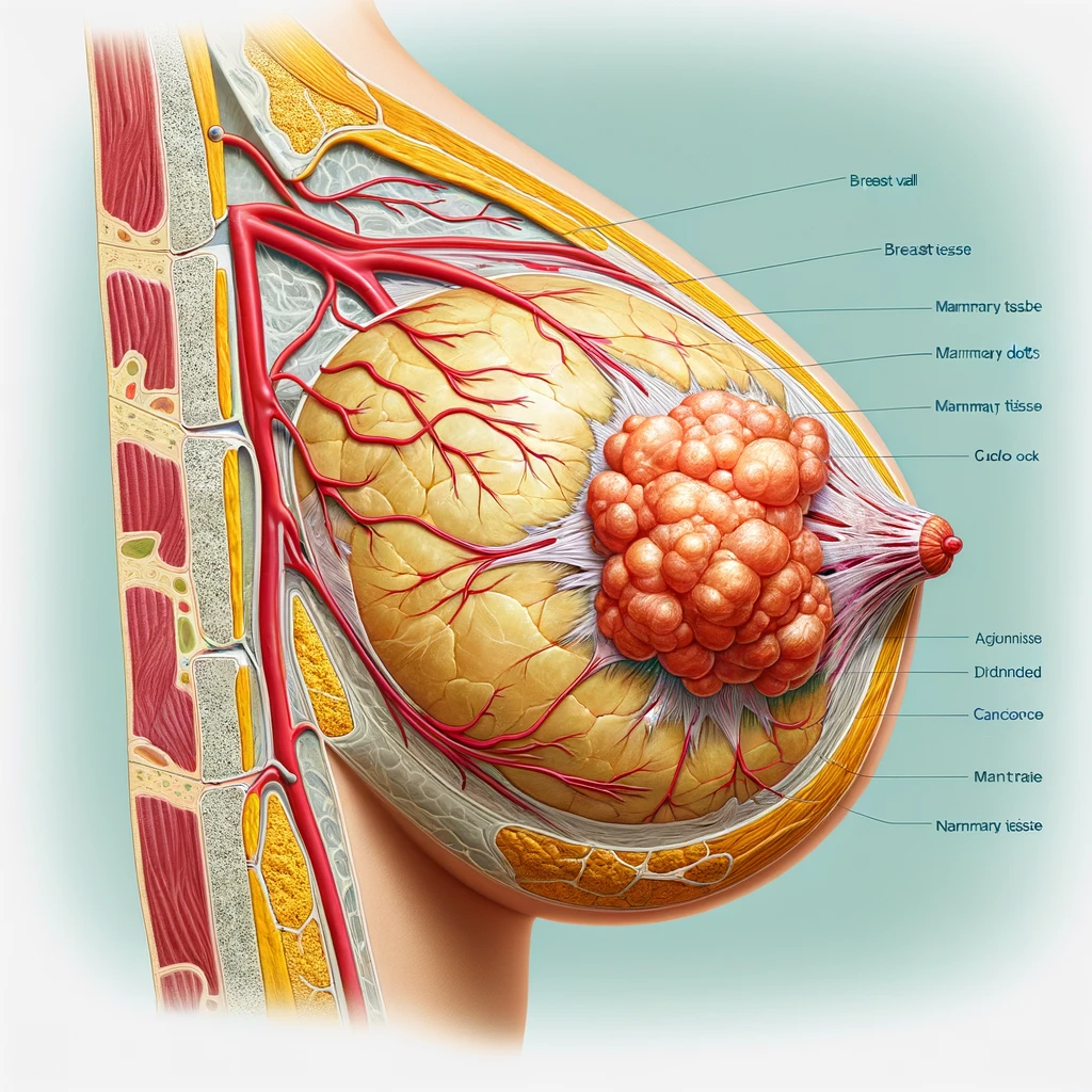
Benign breast tumors are neoplasms that do not infiltrate surrounding tissue, metastasize, or lead to systemic adverse effects. They most often develop from the epithelium, but may also originate from other breast tissues.
About the disease
It’s important to recognize that certain benign conditions can potentially turn malignant. Every tumor carries a specific risk of becoming cancerous. For some, this risk is minimal or virtually non-existent, necessitating only regular monitoring. However, for others, the likelihood of cancer development is high, demanding proactive measures, possibly including surgical removal of the affected tissues.
Benign breast tumors in women can vary in their presentation. Some may be symptomless, detected only through imaging techniques, while others might cause breast discomfort, pain, or unusual nipple discharge. These symptoms warrant further instrumental diagnostic procedures to identify any localized growths.
The primary diagnostic tools for breast tumors are ultrasound scanning and X-ray mammography. Ultrasound is typically recommended for younger women during initial examinations, while mammography is advised for women over 40. When abnormalities are found, both methods are used complementarily.
The necessity of surgery for benign breast tumors hinges on their morphological characteristics, the patient’s individual cancer risk, and clinical symptoms. Tumors with a high potential for malignancy often require preemptive surgical intervention, such as in the case of intraductal papillomas. Conversely, benign tumors like lipomas, which have a low risk of malignancy and are asymptomatic, are usually not treated but monitored by a mammologist. Each case is treated uniquely, with a personalized management plan focused on overall health recovery and cancer prevention.
Types of benign breast tumors
According to the histological sign, the following types of benign breast tumors are distinguished:
- A fibroadenoma is an epithelial tumor with connective tissue components;
- An adenoma is an epithelial tumor of mammary tissue;
- Lipoma is a benign tumor that develops from fat cells;
- A leaflet-shaped tumor is a rare fibroadenoma that develops from the glandular tissue of the breast and is associated with a high likelihood of malignant degeneration;
- Hamartoma – a tumor-like lesion of the encapsulated nodule type that involves different tissue components of the mammary tissue;
- Atheroma – a cyst that forms when the outlet duct of the sebaceous gland is blocked;
- An oleogranuloma is a focal necrosis of fatty tissue that can develop as a result of trauma, inflammation, or surgery;
- Solitary cyst of the breast, associated with the expansion of the lobule against the background of obstruction of the diversion duct;
- Intraductal papilloma is a localized overgrowth of the epithelial and stromal component of the ducts inside their lumen;
- A nipple adenoma is a papilloma that is located inside the milky ducts.
According to histological classification, all benign breast tumors fall into 2 categories from a practical point of view:
- neoplasms without atypical changes;
- neoplasms accompanied by the appearance of cellular atypia.
Symptoms of a benign breast tumor
The main symptoms of a benign breast tumor may include:
- discomfort and painful sensations in the area of the affected breast – in women with a preserved menstrual cycle, the pain can be cyclic (observed in the second phase on the eve of menstruation and passes with its onset) or acyclic (occurring out of connection with the menstrual day);
- local thickening in the breast, which, as a rule, is found in the area of tumor localization;
- discharge from the nipple, which may vary in appearance (from transparent to dark hemorrhagic).
Some tumors are silent and are accidentally detected during preventive examinations.
Features of clinical symptomatology directly depend on the morphological characteristics of the neoplasm. Thus, intraductal papillomas are often manifested by bloody and brown discharge from the milky ducts. In fibroadenomas and adenomas, pain syndrome is a common phenomenon. In oleogranulomas, a localized thickening of the glandular tissue is defined, which may be fused with the surrounding tissue and skin, which creates difficulties in the differential diagnosis with respect to breast cancer. Therefore, patients with local changes detected on ultrasound or mammography are prescribed an extended examination program.
Causes of benign breast tumors
The true causes of benign breast tumors cannot be identified in all clinical cases. The most important risk factors may be:
- psycho-emotional stresses associated with dissatisfaction with the family situation, sexual life, conflict situations at home and at work (these circumstances can disrupt the neurohumoral regulation of the mammary gland);
- unhealthy foods – frequent overeating of animal fats, abuse of trans fats, simple carbohydrates, lack of vegetables, fruits, vitamins, minerals and crude fiber in the diet;
- unfavorable reproductive characteristics – early onset of menstrual function and late cessation of menstrual function, absence or 1 childbirth in the anamnesis, late age of the first childbearing (35 years and older), delivery of a large fetus (weight more than 4 kg), a large number of pregnancy terminations, a short lactation period or its complete absence;
- disturbed functional state of the ovaries, which leads to hormonal imbalance (primarily a relative or absolute deficiency of progesterone);
- ovarian inflammatory diseases secondary to triggering functional disorders;
- diseases of genital organs associated with estrogen imbalance (uterine myoma, endometrioid pathology, hyperplastic processes of uterine mucosa);
- diseases of the thyroid gland, accompanied by a decrease in its functional activity;
- congenital dysfunction of the adrenal cortex;
- excessive prolactin in the body;
- diabetes and insulin resistance;
- obesity and the associated chronic inflammatory response of the body;
- insufficient functional activity of the liver;
- genetic disorders – carrying mutant genes BRCA-1, BRCA-2.
The combination of several adverse factors increases the risks of developing benign breast tumors, some of which may even lead to malignancy over time.
Diagnosis of benign breast tumors
According to clinical guidelines, the generally accepted diagnosis of benign breast tumors includes the following methods:
- clinical examination and palpation of the chest;
- X-ray mammography;
- a breast ultrasound scan;
- Diagnostic puncture and histologic examination, which is indicated at high cancer risk.
In complex clinical cases, a CT scan, magnetic resonance imaging or positron emission tomography is considered.
If a malignant volumetric process in the breast is suspected, an ultrasound-guided puncture biopsy is performed. The obtained material is subjected not only to histologic but also to immunohistochemical (IHC) examination. IHC analysis allows to determine the presence of relevant proteins, including receptor proteins, on tumor cells, which is important for the selection of the most rational treatment tactics.
Treatment of benign breast tumor
Treatment of benign breast tumors depends on many factors. In some cases, dynamic observation is carried out, in others – conservative therapy is prescribed, in others – surgical intervention is indicated.
Conservative treatment
Conservative therapy is aimed at improving the receptor perception of hormones by breast structures, optimizing metabolic processes and correcting predisposing factors. Some patients are recommended to take hormonal drugs as part of complex therapy.
Surgical treatment according to clinical guidelines
Surgery for benign tumors may be recommended in the following cases:
- proliferative, atypical fibroadenoma;
- large size of the neoplasm;
- tumors associated with increased oncological risk (e.g., intraductal papillomas, Phyllodes tumors);
- symptomatic forms of the disease (pain syndrome).
The standard scope of surgery is the removal of the pathologic formation within healthy tissues. The removed material is necessarily sent for histological diagnosis to exclude malignant transformations.
All these types of surgery are performed in more than 750 hospitals worldwide (https://doctor.global/results/diseases/benign-breast-tumor). For example, lumpectomy can be done in 33 clinic across Germany (https://doctor.global/results/europe/germany/all-cities/all-specializations/procedures/lumpectomy).
Prevention
The following interventions are useful in reducing the risk of benign breast disease:
- control of the menstrual cycle and regular preventive check-ups with a gynecologist;
- palpation and self-examination of the mammary glands after the end of menstruation;
- avoidance of smoking and excessive alcohol consumption;
- a balanced, rational diet enriched with vitamins, minerals and omega-3 fatty acids;
- sufficient fiber intake (the recommended amount per day is 30 g);
- optimal water balance (drinking about 2 liters of pure water per day);
- rational alternation of work and rest;
- Creating a favorable psychological climate in the family and at work.
Rehabilitation
Surgical interventions performed for benign breast tumors are minimally traumatic. Therefore, they are characterized by a short period of rehabilitation. During the recovery phase after tumor removal, it is recommended to maintain a moderate level of physical activity, follow a healthy lifestyle and regularly visit a mammologist.