Brain aneurysm
What is a brain aneurysm?
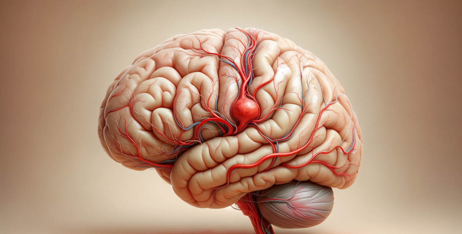
A cerebral aneurysm is a pathological localized bulge in the thinned wall of an artery. It is a “time bomb” that “explodes” at least once in a person’s life – the wall bulge bursts, and blood contained in the aneurysm flows to the brain. Such a vascular catastrophe can cause permanent neurological damage and even endanger the patient’s life.
If symptoms of this condition occur, the patient should see a doctor as soon as possible to give them specialist medical attention. The ideal scenario is identifying and blocking the aneurysm from the bloodstream before it breaks down. It is possible if the person is vigilant about their health – timely treatment by a qualified neurologist in the presence of complaints of headaches or other neurological disorders.
Disease
Aneurysms more often affect the vessels within which blood flows under high pressure – the area of their bends or the areas of the bifurcation. Thinning of the wall and high pressure leads to an increase in the vessels’ diameter, forming pathological aneurysms. Gradually, it begins to compress the surrounding brain structures, disrupting the functions of the central nervous system.
The pathology described is dangerous for the patient because of the high risk of rupture of the aneurysm with bleeding inside the skull. Ruptured aneurysms are more common in women at least 50-65 years of age.
Types of cerebrovascular aneurysms
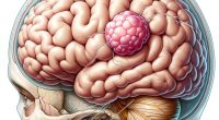
The classification of cerebrovascular aneurysms is based on several criteria. Depending on the shape, the protrusion may be bag-shaped or spindle-shaped. The first type of aneurysm is more common.
Depending on the size of the protrusion, there are:
- miliary (up to 3 mm in diameter);
- regular size (up to 15 mm);
- large (up to 25 mm);
- giant (over 25 mm).
The larger the bulge, the more it compresses the brain tissue and causes a more intense neurological image. Protrusions can also be single- or multi-chambered, single or multiple, congenital or acquired.
Symptoms of cerebral vascular aneurysm
The danger of a bulging arterial wall is their “silent” development. In the early stages, the patient may not even know of a severe problem because small aneurysms practically do not pinch brain tissue and do not cause unpleasant sensations.
The clinical picture emerges in the later stages of the disease. The first signals that there may be a problem are:
- persistent headaches in a particular area;
- vision loss, pain in the eyes and head;
- changes in the motor function of the limbs (the patient may suddenly “forget” how to hold a spoon, or their handwriting suddenly deteriorates);
- breaks the tactile sensation and pain sensitivity in the feet;
- seizures;
- numbness of facial muscles, inability to smile, sudden loss of vocabulary.
These signs suggest a compression of brain tissue. A neurological deficit develops that should promptly prompt you to seek help from a neurologist.
If the disease progresses, the aneurysm may rupture. This condition is already directly threatening the patient’s life. Symptoms of rupture include:
- sudden severe headache that does not respond well to medication correction;
- photophobia;
- nausea and vomiting;
- double vision;
- loss of consciousness;
- severe cramps.
You need to call an ambulance team if the person has described symptoms.
Causes of brain aneurysm
A vascular aneurysm in the brain can be congenital or acquired. In the first case, the disease occurs against the background of a pre-existing defect in the development of one of the artery walls. It may damage the vessel’s inner lining by forming an aneurysm at an early age, which is one of the manifestations of several diseases (Marfan syndrome, Randu-Osler, Ehlers-Danlos).
Acquired aneurysms can be caused by:
- head and brain injuries;
- hypertension;
- infections;
- cancer of the intracranial structures;
- addiction to drugs;
- atherosclerotic lesion on the inner surface of the blood vessels.
The provoking factors of the disease are constant stress and heavy physical exertion. They cause an increase in pressure on the vessels in the head, increasing the risk of mechanical damage.
Diagnosis of brain aneurysm
Detecting abnormalities in brain arteries is not always an easy task, especially if the patient does not feel any pain. In such cases, the disease is detected incidentally during a preventive examination or investigation because of suspicion of another pathology.
To diagnose an aneurysm, if the patient has complaints and objective symptoms of a neurological deficit, the neurologist will prescribe the following instrumental diagnostic methods:
- computed tomography or CT scan procedure is often used to determine an already existing rupture of the vessel where there has been bleeding into the brain tissue;
- magnetic resonance imaging (MRI);
- angiography of brain vessels – a method that allows you to assess the size, location, and structural characteristics of a pathological bulge.
If subarachnoid hemorrhage is suspected because of a ruptured aneurysm, a neurologist will perform a lumbar puncture to assess the composition of the cerebrospinal fluid.
Treatment methods for cerebrovascular aneurysms
The management of patients with intracranial artery disease is always complex. The neurologist will assess the risk factors that mainly affect the patient. Based on the information obtained, the specialist prescribes medications that influence the mechanisms of development of secondary aneurysms and promote stabilization of the person’s condition.
To prevent the aneurysm from rupturing, the patient is advised radical treatment surgery. Emergency intervention is indicated for aneurysm rupture.
Conservative treatment
In this area, the most commonly used methods include:
- painkillers;
- blood pressure medicines to control blood pressure;
- antiepileptic drugs;
- means reducing the concentration of cholesterol in the blood.
Conservative treatment complements surgery and helps prevent relapses in the presence of risk factors.
Surgical treatment of cerebral vascular aneurysm
The best way to treat an aneurysm is to cease the blood supply with a surgical intervention. It eliminates ruptures and adverse effects on the central nervous system. The preferred method is endovascular surgery. The neurosurgeon punctures the femoral artery, inserts a thin wire (an introducer) and directly accesses the vessel with the aneurysm, and then, using miniature instruments, fills the aneurysm with platinum micro-thread or installs a stent, which strengthens the blood vessel wall in the damaged area of the artery, after which it removes the instrumentation from the arteries. Not all manipulation is done blindly – the doctor notes what is happening through the screen of a particular X-ray machine.
Less frequently, open brain interventions are used. A neurosurgeon treats the trepanation of the skull, dissects the meninges, reaches the blood vessel affected by the aneurysm, and uses micro-tools to perform the necessary manipulations – excising the protrusions, tying them on both sides or clotting them, then sutures the damaged tissues and closes the trepanation with the patient’s bone. The size of this hole varies greatly depending on the clinical situation. The choice of a particular treatment option depends on the individual characteristics of each clinical case.
All these surgical procedures are performed in 701 hospital worldwide (https://doctor.global/results/diseases/brain-vessel-aneurysm). For example, you can have an endovascular coiling for a brain aneurysm in 28 clinics in Turkey with approximate cost of $15 K. (https://doctor.global/results/asia/turkey/all-cities/all-specializations/procedures/endovascular-coiling-for-brain-aneurysms).
Prevention of brain aneurysm
There is no specific prevention of aneurysms. It is recommended to monitor blood pressure, regulate cholesterol and blood sugar, and avoid stressful factors and physical overexertion.
Patients whose relatives have been affected by a similar problem should regularly undergo comprehensive neurological examinations for early detection of pathology.
Rehabilitation after surgery
The recovery time after surgery on the brain aneurysm is individual for each patient. Depending on the severity of the neurological damage, patients may stay in the hospital for several days to weeks after surgery. A neurosurgeon will perform daily dressing care, monitoring the person’s well-being.
If the persistent neurological deficit persists after treatment, an individualized rehabilitation plan is selected for the patient. It may include therapeutic massage, physical therapy, medication, spa treatment, and physical therapy.
Advances in Brain Aneurysm Research
Recent advances in medical research have led to improved surgical techniques, better diagnostic tools, and a deeper understanding of the biological mechanisms behind aneurysm formation and rupture. Ongoing studies continue to explore genetic factors and innovative treatments.
Conclusion
A brain aneurysm represents a significant medical challenge, but with advances in medical science, many patients lead full and active lives post-diagnosis. Awareness, early detection, and modern treatment strategies are key to effectively managing this condition. Continued research and patient education remain critical in further improving outcomes for those affected by brain aneurysms.