Chest wall deformity (Pectus excavatum)
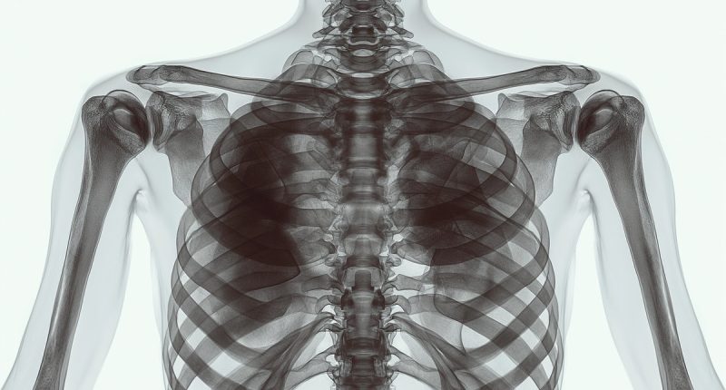
Pectus excavatum is an anomaly in which the sternum and the front ends of the ribs are pressed inward. This deformity is dangerous because it increases mechanical pressure on the lungs and heart muscle, leading to impaired function.
Disease
Among malformations of the chest, pectus excavatum is the most common. It accounts for about 90% of cases. Boys are the most vulnerable – they are five times more likely to have this disease.
The leading cause of this disease is excessive growth of cartilage connecting the sternum and ribs. It occurs when bone growth tissue lags behind the development of cartilage. As a result, the sternum appears to be prolapsed into the chest cavity. In some cases, the deformity may have an acquired origin.
In 80% of children, the disease manifests itself in the first year of life, but progression occurs during the maximum growth of the child: 6-8 and 11-13 years. Symptoms of chest wall deformity include not only a visible deformity but also clinical signs of impaired heart and lung function. The latter becomes particularly pronounced in adult women and men, associated with the progression of the malformation.
How to correct a chest deformity? The choice of treatment method is determined by the degree of deformity. In the early stages, when there are no negative consequences, conservative treatment is possible. With pronounced curvature, when the functional state of the heart and/or lungs is affected, the question of surgical correction is decided.
Different forms
According to the description of the external shape of the chest, there are two types:
- symmetrical deformation.
- asymmetric deformations.
The severity of the pectus excavatum is estimated by the Haller index. It is calculated by dividing the transverse diameter of the chest by the anterior-posterior distance on CT of the chest on the axial slice, which demonstrates the smallest distance between the anterior surface of the vertebral body and the posterior surface of the sternum. These distances are measured from a chest CT scan.
The following values are used:
- normal chest: <2.0;
- mild excavatum: 2.0-3.2;
- moderate excavatum: 3.2-3.5;
- severe excavatum: >3.5.
Corrective pectus excavatum surgery is considered with a Haller index ≥3.25.
Symptoms of a chest wall deformity
Along with the complaints of the curvature of the chest, which constitutes several complexes and psychological problems in adolescents, there are many symptoms associated with secondary changes in the organs located in the chest cavity. It is due to chronic compression, as well as due to dysplasia of connective tissue components, which affects not only cartilage and ribs but also lung tissue, contributing to the formation of bronchiectasis (local enlargement of bronchi) and broncho-obstructive syndrome. Congenital malformation of the chest is characterized by “paradoxical breathing,” which manifests as retraction of the sternum and ribs with inhalation. As a result, patients often suffer from respiratory diseases over a prolonged course. Compression of the lungs, difficulty breathing, sputum accumulation in the bronchi leading to shortness of breath and coughing.
Compression of the ribs and sternum of the heart increases the load on the right ventricle. Conduction of nerve impulses is disrupted, resulting in heart rhythm disorders. Patients are concerned about “palpitations” at rest, mild fatigue with light exercise, pain on the left side of the sternum, shortness of breath, and increased blood pressure.
Reasons for pectus excavatum
The causes of chest wall deformities have been formulated through various theories.
- Genetic theory. Close relatives have similar anatomical changes. This theory explains the occurrence of the malformation by mutations in the genes that regulate the collagen fibers that make up connective tissue. It is associated with a breakdown in the synchronous growth of the bone-chest compartment.
- Acquired theory. Chest wall deformity can form after injuries, burns, and cuts. These causes alter the normal shape of the breast and lead to its curvature. Acquired deformity can also result from tuberculosis infection, osteomyelitis of the rib bones, lateral curvature of the spine, and rickets.
Diagnosis of pectus excavatum
According to clinical guidelines, the diagnosis of a pectus excavatum includes the following methods:
- a clinical examination;
- laboratory tests;
- X-ray or CT scan of the chest;
- EKG;
- Ultrasound of the heart with Doppler;
- Holter monitor;
- a study of respiratory function in the lungs.
Treatment chest wall deformities
The only radical way to treat pectus excavatum is to perform surgery.
Conservative treatment
Conservative methods may be effective only for first-degree deformation. They aim to strengthen the muscle corset and improve the functional status of the lungs and heart. Physiotherapy, swimming, massage, and wearing a special corset are indicated.
With the progression of chest deformity, more permanent changes are formed, as can be seen in the EKG, hypertrophy of the right heart is more often observed, as well as metabolic changes in the heart muscle. Patients may experience tachycardia or bradycardia, His bundle block, or hypertrophy of the right heart. In this situation, management must be performed not only by an orthopedic traumatologist but also by a cardiologist. If respiratory diseases develop, the pulmonologist is also involved in providing health care.
Surgical treatment
Surgical correction is performed for II-III degrees of thoracic curvature.
Since 1998, the minimally invasive method of thoracoplasty, according to D. Nuss, has been widely used. This method is based on “extensions” of the rib cartilage with the help of a metal plate located behind the sternum. This method does not require partial removal of rib cartilage and dissection of the sternum. The plate installation is performed through small entrances, which reduces trauma to the chest muscles, reduces blood loss during surgery, and has a significant cosmetic effect.
This diseased is treated in more than 530 hospitals worldwide (https://doctor.global/results/diseases/chest-wall-deformity). For example, chest wall deformity surgery is performed in 27 clinics across Turkey for approximate price of $5.7 K. (https://doctor.global/results/asia/turkey/all-cities/all-specializations/procedures/chest-wall-deformity-correction)
Prevention
Prevention of acquired forms of the disease is based on preventing thoracic injuries.
Rehabilitation services
After surgery, because of the restoration of the shape of the chest, the heart takes a more physiological position; there is no compression of the right parts of the heart, which contributes to changing the valve position of the structures. After surgical treatment, patients also notice an improvement in well-being and the ability to tolerate heavy physical exertion. At that point, rehabilitation and individually selected physical exercises are helpful.
Conclusion
Pectus excavatum is a congenital chest wall deformity that affects individuals both physically and emotionally. While it can pose challenges, various treatment options are available to address the condition’s severity and improve the quality of life for those living with it. Understanding the causes, symptoms, diagnosis, and treatment options for pectus excavatum is essential for individuals and their families seeking information and support.


