
Forearm fracture
What’s that?
Forearm bone fractures are a type of injury in which the bone integrity of either the radius or ulna (sometimes both bones at once) is compromised.
About the disease
According to different authors, forearm fractures are common, accounting for 25.3% to 27.3% of the total number of fractures. The radius’ distal end is most often fractured in older adults. That is followed by fractures of both bones of the forearm. An isolated ulna fracture is less common than a fracture of both bones.
This type of injury is manifested by swelling and lividity of soft tissues in the area of injury. The patient is bothered by intense pain, which increases when trying to move the arm, as well as when palpating. With intra-articular fractures, blood can accumulate between the articulating bones, which subsequently creates conditions for the development of deforming osteoarthritis. Fractures of the forearm bones can be open when there is a skin wound and closed when the soft tissues are intact.
Treatment of forearm bone fractures can be carried out by both conservative and surgical methods. The optimal patient management plan is made by a traumatologist, considering the injury’s peculiarities and the person’s professional activity.
Types
According to the classification, the following groups of traumatic injury to the bony base of the forearm are distinguished:
- in intra-articular fractures in the elbow joint, which can be either without displacement of bone fragments or with displacement:
- violation of the bone integrity of the ulna;
- violation of bone integrity in the area of the head and neck of the radius;
- violation of the integrity of the ulnar coronoid process, which may be combined with its detachment.
- fractures in the body of one or two bones of the forearm, which may be displaced or non-displaced
- disruption of the integrity of two bones;
- isolated injury to the ulnar bone, which may be accompanied by dislocation of the head of the radius;
- dislocation of the ulnar head may accompany an isolated injury to the radius.
- disruption of the integrity of the lower end of the radius (fractures can be displaced and non-displaced):
- typical epiphyseal fracture and epiphysiolysis with or without detachment of the ulnar spinous process;
- fractures of the lower segment of the radius may be combined with a similar injury to the ulna.
Eponyms of forearm fractures fall into a separate category:
- Monteggia fracture is a disruption of the integrity of the ulna combined with dislocation of the head of the radius at the elbow joint;
- Galeazzi fracture – violation of the integrity of the radius in the lower part combined with dislocation of the head of the ulna in the area of the wrist joint.
Symptoms of fractured forearm bones
Symptoms of fractured forearm bones include the following signs:
- marked swelling of the tissues, which appears shortly after the injury;
- spread lividity of the skin and subcutaneous tissue associated with traumatic damage to small feeding vessels;
- intense pain due to irritation of nerve endings;
- impaired functional activity of the hand (inability to move the fingers, bend-extend the elbow, etc.);
- presence of a skin wound (in open fractures);
- soreness when you feel the forearm;
- inability to hold objects with your hand;
- change in the appearance of the hand, the appearance of pathological curvature;
- presence of unnatural mobility in the forearm (in the case of a complete fracture of both bones);
- crunching due to displacement of bone fragments.
When an injury occurs in the elbow area, the arm hangs like a whip.
Reasons
The causes of broken bones in the right or left forearm can be as follows:
- fall on the elbow joint, which can be either bent or unbent;
- direct punch to the elbow or wrist or the middle part of the forearm;
- fall with a hand support.
Fractures can result from sports, street and occupational injuries.
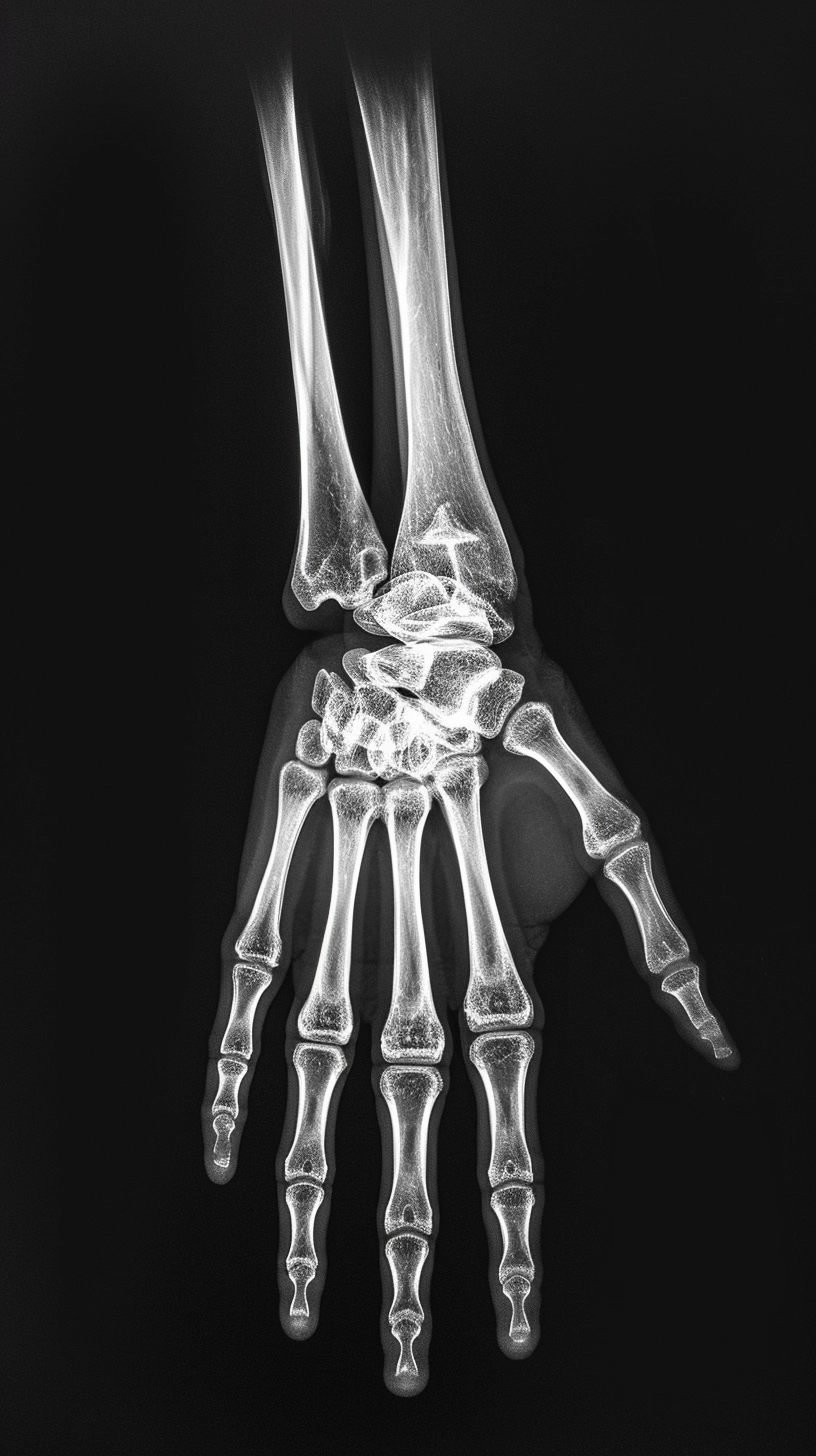
Diagnosis
In diagnosing forearm fractures, the doctor is guided by objective examination results and X-ray scanning. X-rays are performed in at least two projections and allow for visualization of the fracture line, which looks darker, and assessing the position of bone fragments. These data are necessary for planning the optimal treatment program. Thus, if bone fragments are displaced by more than 0.5 cm, the indications for surgical intervention expand.
If a joint injury is suspected, arthroscopy, a diagnostic examination of the joint cavity from the inside, may be performed. During this procedure, a magnified image is displayed on a monitor screen. If necessary, diagnostic arthroscopy can be converted into treatment, which consists of removing foreign objects from the joint, removing clots, and washing the intra-articular cavity.
Treatment of fractured forearm bones
The characteristics of the injury determine the specifics of the treatment program, the patient’s age, and occupation. Treatment of fractures can be carried out by both conservative and operative methods.
Conservative treatment
Conservative tactics are applicable when there is no displacement of bone fragments. It consists of applying a circular plaster cast, which provides reliable immobilization of the bones. In the presence of displaced fractures, conservative treatment is not acceptable because the incidence of such complications as contractures, false joints (nonunion of bone fragments with preservation of pathological mobility between them), posttraumatic club hand, etc., increases.
Skeletal traction may be performed to align the bone fragments. By applying gradual pressure in a predetermined direction, the doctor helps to create the correct position of the forearm bones. Once the planned result has been achieved, a plaster cast is applied to the arm with the above- and below-lying joints captured.
Surgical treatment
Surgery for forearm bone fracture is performed in complicated injuries, including those accompanied by dislocation of bone fragments. The latter are repositioned by an open access, after which fixation is performed. It can be performed in a periosteal way (plates and screws) or intraosseous method (fixation spokes are inserted inside the bone). Periosteal osteosynthesis is the most acceptable, as it involves fewer complications.
All these treatment options are available in more than 800 hospitals worldwide (https://doctor.global/results/diseases/forearm-fracture). For example, forearm fracture surgery can be done in 40 clinics across Germany for an approximate price of $7.3 K (https://doctor.global/results/europe/germany/all-cities/all-specializations/procedures/forearm-fracture-surgery).
Prevention
Use extra caution during icy weather to reduce the risk of forearm bone fractures. It is important to remember safety rules when performing household chores and sports activities according to proper technique.
Rehabilitation
After the plaster cast is removed, the active rehabilitation phase begins, which consists of the following areas:
- performing physical therapy exercises aimed at developing the joint;
- taping;
- physiotherapy treatments;
- a course of therapeutic massage.
The average length of the rehabilitation period is 2-4 weeks.