Double outlet right ventricle (DORV)
Determination
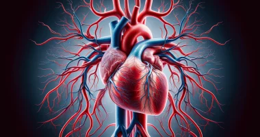
Double outlet from the right ventricle (DORV), (partial transposition, Taussig-Bing anomaly), is a group of congenital heart defects, more often caused by genetic anomalies, characterized by an abnormal type of ventriculo-arterial connection, in which the aorta and pulmonary trunk originate completely or predominantly from the right ventricle. This malformation occurs due to impaired rotation of the arterial conus and its wedging between the atrioventricular valves and is accompanied by impaired formation of the outlet from the left ventricle.
Epidemiology
Clinically, the incidence of this malformation is 0.7%, which is quite rare, but according to data from autopsies, it is 2.7%, which indicates a low detection rate and early mortality of persons with this disease. If we take all congenital heart defects, only 1.5% of patients undergo surgical correction of this defect, with an average mortality rate of almost 10%. The prognosis of the course of the malformation depends mainly on its type and, accordingly, the hemodynamic variant.
Life expectancy is higher in patients with a hemodynamically more favorable course, namely, right ventricular outflow tract obstruction. In general, the time during which 50% of patients with this anomaly die does not exceed five years, and only 17% of patients with all types of malformation live to 15 years.
Classification
– Tetralogy of Fallot type DORV. In this case, the ventricle septal defect (VSD) is located subaortically or subarterially, combined with right ventricular outflow tract obstruction;
– VSD DORV type. It is characterized by the same, but in the absence of right ventricular outflow tract obstruction;
– Transposition of the great vessels DORV type. It is characterized by the absence of right ventricular outflow tract obstruction, and the VSD is located subpulmonary;
– DORV with non-commitment VSD. It is manifested by a VSD in the sinus or trabecular septum and may be combined with right ventricular outflow tract obstruction.
Diagnosis
The diagnosis of this disease is based on the characteristic symptoms of heart failure, which manifest from birth. The gold standard for detecting this malformation is echocardiography, which allows the assessment of the anatomical structure of the heart and its hemodynamics. Prenatal diagnosis is possible, but it is very difficult to perform.
Echocardiography most commonly reveals a large, high interventricular septal defect, pulmonary valve insufficiency, concentric marked hypertrophy of the right ventricular myocardium, moderate dilatation of the right atrium, and significant dilatation of the pulmonary artery trunk and branches, along with marked pulmonary hypertension.
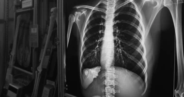
According to chest radiography, the expansion of the heart shadow to the left and signs of hypervolemia in the small circle of blood circulation are noted.
Treatment
Diuretics, ACE inhibitors, cardiac glycosides, β-blockers, and catecholamines are recommended for patients with DORVat the stage of preoperative preparation.
β-blockers and benzodiazepines for hypoxic attacks are recommended in patients with DORV of the Fallot’s tetralogytype. Calcium antagonists, endothelin receptor blockers, phosphodiesterase type 5 inhibitors, and prostaglandin analogs are recommended for treating pulmonary hypertension in patients with DORV.
The size of the left ventricle determines the type of surgical correction and, accordingly, the localization of the ventricular septal defect. Many patients undergo palliative surgery before reconstructive correction of this malformation, especially if the left ventricle is hypoplastic or borderline in size.
Palliative surgeries include the Blalock-Taussig shunt (anastomosis between the subclavian and pulmonary arteries), pulmonary artery banding (pulmonary artery narrowing), and sometimes a resection of the coarctation of the aorta or the first stage of the Norwood procedure (correction of hemodynamics by reducing the burden on the pulmonary artery caused by increased blood flow).
Taking into account that obstructive pulmonary vascular damage develops as rapidly as in VSD, patients with DORVshould be operated on early in infancy (before six months of age). Early age should not be considered a factor in hospital mortality. The preferred surgery method is to create an intraventricular tunnel with a patch of dacron or Gortex tissue connecting the LV to the aorta. Some children undergo pulmonary artery banding beforehand, but this should not be considered a rule – early correction of the malformation is much more critical.
In the case of a restrictive ventricular septal defect (when the size is less than the diameter of the aortic valve), it is widened by an anteroseptal incision or by excision of the interventricular ridge in this area. Muscle bundles obstructing the outflow tract are excised to form a direct tunnel between the aorta and the VSD.
All these treatment options are available in more than 320 hospitals worldwide (https://doctor.global/results/diseases/double-outlet-right-ventricle-dorv). For example, Glenn procedure can be done in 28 clinics across Turkey for an approximate price of $20.5 K (https://doctor.global/results/asia/turkey/all-cities/all-specializations/procedures/glenn-procedure).
Conclusion
The incidence of this malformation is very rare. According to some authors, this anomaly occurs in 2-3% of children born with congenital heart disease. However, the rarity of the occurrence of this malformation does not exclude the likelihood of meeting with it. It should be remembered that this pathology requires surgical treatment at an early age (up to 6 months), and drug therapy is supportive and only distances the outcome of inevitably increasing chronic heart failure.


