
Fractured finger
What’s that?
A finger fracture is a traumatic injury to the bony tissue of either phalanx.
About the disease
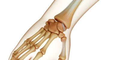
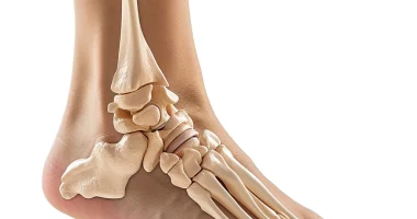
Traumatic injury to the finger phalanges is possible on both the hand and foot. In turn, each finger consists of 3 phalangeal bones:
- the furthest distal one;
- close to the metacarpal (on the hand) or metatarsal (on the foot) proximal;
- the middle one in between.
However, there is an exception to this rule – the thumb. Its anatomy is represented by only two phalanges – proximal and distal. In some people, the little finger can also be bi-phalangeal, which is considered a variant of the norm.
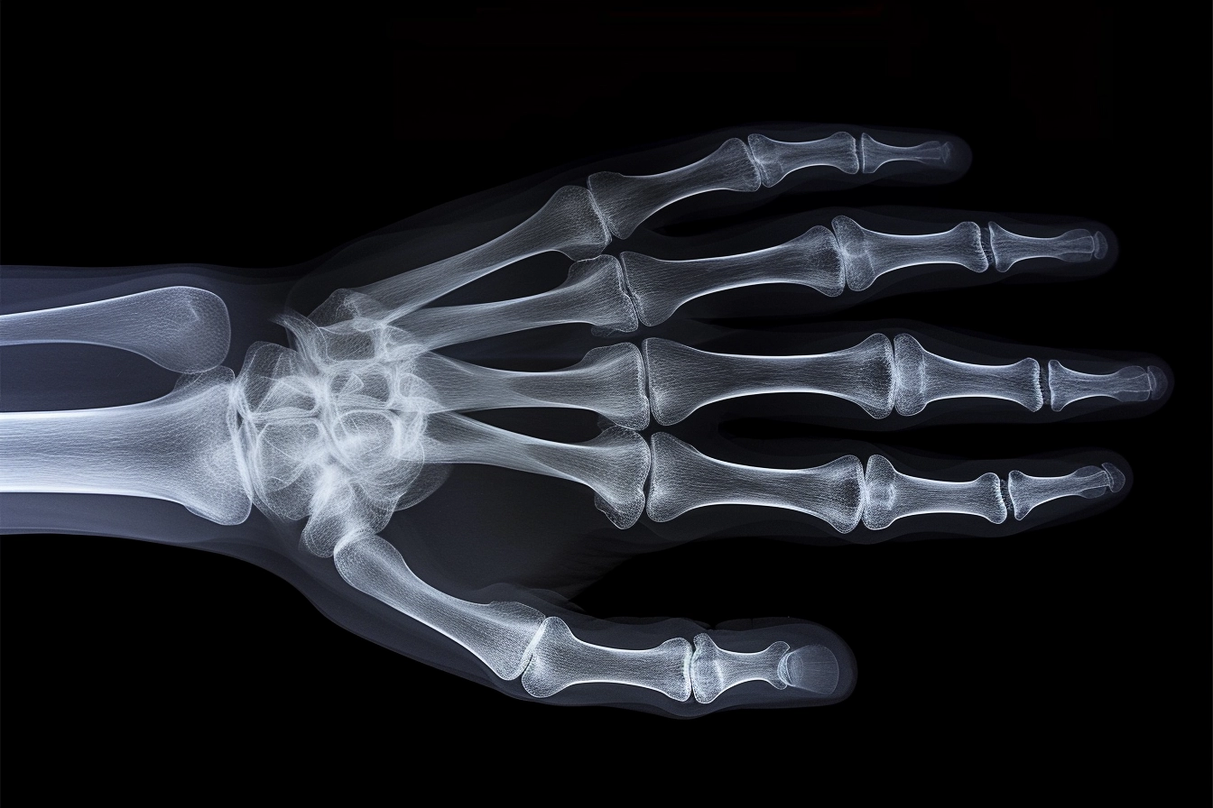
The causes of a fractured finger may be related to direct or indirect traumatic injury. This condition is manifested by typical symptoms – intense pain sensations, swelling, and lividity of the injured finger, the presence of a wound (in the case of an open fracture), deformation of the finger, inability to bend-extend the phalanx and pathological mobility. Based on clinical symptoms, the doctor establishes a preliminary diagnosis, which is confirmed by radiography.
In most cases, treatment of a finger fracture is carried out by conservative methods. Only in the case of complicated injuries is the question decided in favor of surgical intervention.
Types of finger fractures
The types of finger fractures distinguished in practical traumatology take into account different classification criteria:
- open and closed injuries, the difference between which lies in the intactness of the skin (the presence of a skin wound is characteristic of an open fracture);
- isolated and combined injuries – in the second case, bone damage is combined with damage to other structures;
- oblique, transverse, spiral, and other varieties that are defined by the visual characteristics of the fracture line;
- injuries in which bone fragments retain the typical bone direction or are characterized by displacement.
Types of fractures of the finger of the hand
A particular type of finger fracture is a mallet fracture (Busch fracture, Segond fracture). This term refers to a particular type of fracture of the distal phalanx of the finger. In this injury, a triangular bone fragment is detached from the base of the terminal phalanx along with the tendon of the finger extensor muscle. This fracture occurs due to unnatural flexion of the finger, which is in an extended state. The finger takes the shape of a hammer, and active extension becomes impossible.
Most modern surgeons use the J.R. Doyle classification when discussing mallet finger injuries:
- Type 1 is a closed injury in which the extensor tendon component is damaged at the level of the last interphalangeal joint, and a small bone fragment may be detached from the distal phalanx;
- Type 2 – open injury to the extensor tendon component at or just above the level of the last interphalangeal joint;
- Type 3 – open wound with damage not only to the skin but also to the underlying soft tissues, as well as damage to the extensor tendon component at the level of the last interphalangeal joint or slightly above;
- Type 4 – the base of the distal phalanx is broken, which may be combined with subluxation of the phalanx.
Symptoms of a fractured finger
The brightness of the clinical picture characterizes the symptoms of a fractured finger. At the moment of injury, an intense pain syndrome appears, which increases with the slightest attempt to move the finger. The traumatized area swells, and soon, there is a livid coloration of the skin (associated with traumatic vascular damage). In the area under the nail, bruising may be detected. When palpating the place of injury, the pain increases significantly, and unnatural mobility and bone crepitation (caused by friction of bone fragments against each other) can be determined. Free movements in the toe become impossible. When the foot is injured, its supporting function is impaired, and when the hand is injured, grasping and holding are affected.
Causes of a fractured finger
The main etiologic factors are:
- A direct damage to the finger with a heavy object (household, industrial, or sports injuries);
- Forced twisting or extension that exceeds the strength of the bone tissue (the distal phalanx is considered the least strong).
Fractures of the finger phalanges are often combined with a skin wound. They occur when fingers are injured at work or home due to non-compliance with safety rules. Bone damage may be combined with injury to the tendon-ligamentous compartment of the finger and dislocation of the finger phalanx.
By etiology, two types of fractures are also distinguished:
- Traumatic, which are caused by high-energy trauma;
- Pathological, in which the integrity of the bone is violated due to its primary damage, for example, against the background of osteoporosis, neoplasm, inflammatory process, etc.
Diagnosis
Objective examination and palpation of the injured fingers allow a preliminary diagnosis. An X-ray scan of the hand or foot is performed for final diagnosis. Radiologic signs of a fracture are:
- bone contour irregularities;
- visualization of the shading line;
- the presence of a curved phalanx.
Treatment of a fractured finger
According to clinical guidelines, phalangeal fractures of the foot or hand are treated with conservative methods. In complex cases, surgical intervention may be required.
Conservative treatment
Conservative treatment is effective when it is possible to restore the fingers’ anatomy accurately using manual techniques. The treatment consists of the following steps:
- anesthetizing the fracture site;
- manual repositioning of bone fragments (in displaced fractures);
- control of the effectiveness of manual repositioning with radiographic examination;
- application of immobilizing rigid dressing (in fractures without displacement, item 2 and item 3 are not carried out).
The duration of immobilization lasts for 2-4 weeks. Immobilization of the bone fragments creates conditions for bone regeneration, which grows in the direction from one fragment to another. The bone callus is fragile at the initial stage, so the bone fragments mustn’t move. Otherwise, it will be traumatized, creating the preconditions for forming a false joint.
Immobilizing dressing covers the entire hand and wrist joint and reaches the middle of the forearm. If the toes are injured, a boot that reaches the middle of the shin is applied.
Surgical treatment
Surgical treatment for fracture of the phalanges of a finger on the hand or foot is performed for multifocal fractures and severe intra-articular injuries. Special screws are used to fix the bone fragments. In some cases, skeletal traction of displaced fragments may be used.
If the flexor or extensor tendons of the fingers are damaged, suturing is indicated. The earlier the operation is performed, the better the functional outcome. If several months have elapsed since the tendon injury, tendon repair is performed.
All these treatment options are available in more than 250 hospitals worldwide (https://doctor.global/results/diseases/finger-fracture). For example, finger/toe amputations is performed in 10 clinics across Germany for an approximate price of $5.9K (https://doctor.global/results/europe/germany/all-cities/all-specializations/procedures/fingertoe-amputation-surgery).
Prevention
Prevention of finger fracture is primarily aimed at preventing occupational injuries – it is recommended to follow safety rules and use personal protective equipment. To prevent domestic injuries, it is essential to exercise caution when gardening, cooking, and other work.
Rehabilitation after a fractured finger
Rehabilitation after a finger fracture is essential for the restoration of fine motor skills of the hand, which are primarily associated not only with the ability to perform household services but also with the performance of professional duties. After removing the plaster cast, massage courses, physical therapy exercises (grasping small objects, etc.), physiotherapy, acupuncture, etc., are performed.