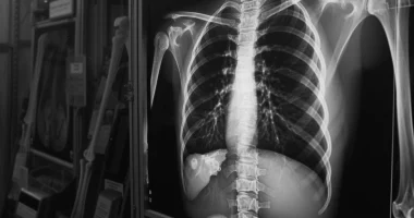
Flat back syndrome
Definition
Flat back syndrome is a pathological condition characterized by decreased expression of one or more physiological curves of the spine. With a flattening of the thoracic curve, there is compression of the heart, accompanied by pain and decreased tolerance to loads. With a decrease in the lumbar curve, the ability to maintain an upright body position is impaired, and patients are forced to bend the joints of the lower extremities during standing and walking. Pain in the back and joints is noted. Pathology is diagnosed by examination, radiography, CT, and MRI. Treatment is predominantly surgical.
General information
Flat back syndrome is a condition in which the angle of thoracic kyphosis is less than 30° (taking into account age, there may be variations from 23° to 40°), and the angle of lumbar lordosis is less than 145°. The congenital variant of the pathology is detected in children and young people, characterized by a decrease in all curves and the presence of cardiac symptomatology. With acquired flat back, middle-aged and older patients are usually affected, the lumbar curve is reduced, and orthopedic disorders are detected.
Causes
Flat back syndrome is a polyetiologic condition. A congenital flat back is a developmental anomaly. Acquired flattening of the spinal column curves is most often formed after surgery on the lumbar spine with the fixation of a metal structure on the sacrum or lower lumbar vertebrae. Other possible causes of flat back development include:
- increased kyphosis at the level of the thoracolumbar junction;
- fractures of the vertebrae of the thoracolumbar spine;
- lumbar spondylosis;
- Vertebral instability with the development of spinal canal stenosis;
- bone block at the thoracolumbar junction;
- ankylosing spondylitis;
- hip contractures.
Symptoms
Congenital form
Smoothing of all spine curves is noted. Cardiologic symptoms usually occur during puberty and often increase with age. Patients complain of decreased stamina, heart pain, and palpitations. Arrhythmias are possible. On auscultation, an ejection murmur is heard, which may be misinterpreted as a murmur of atrial septal defect or pulmonary artery stenosis.
Acquired flat back
Patients with a flat back complain of difficulty standing upright and unsteadiness when walking on uneven surfaces. Externally, the absence or significant reduction of lumbar lordosis is detected. When standing, the body is tilted to the front, the cervical and thoracic regions are overextended, the pelvis is pulled back, and the hip and knee joints are slightly bent.
The lumbar flattening persists with back flexion and attempts at manual correction. Due to constant overstrain, fatigue is noted after minor physical activity, and back pain is common. The pain is usually non-localized, aching, or pulling, predominantly in the thoracolumbar and lumbar region. It increases with prolonged standing and physical exertion.
Due to overextension of the upper spine, the pain syndrome spreads to the neck and upper back. In case of prolonged flat back or primary articular lesion, flexion contractures of the hip joints, less often – knee joints can be determined.
Complications
Reduced endurance in patients with congenital pathology may cause disability. The literature indicates that mitral valve prolapse is often detected in such patients, but it is difficult to establish whether it is a consequence of a flat back or a parallel process. Some researchers mention the compression cardiac loss syndrome, manifested by rhythm and conduction disturbances. Unphysiologic body position contributes to the development of degenerative changes in the vertebral column.
Acquired flat back aggravates the course of the underlying disease of the spine or joints, leads to osteochondrosis, and increases the likelihood of formation of intervertebral hernias, spondyloarthritis, arthrosis of the hip and knee joints, and other degenerative pathologies. Possible compression of spinal roots with the development of neurological symptoms. In severe forms, limitation or loss of ability to work is noted.
Diagnosis
Flat back syndrome is diagnosed by an orthopedic surgeon or neurosurgeon based on objective examination data and the results of additional tests. The history of life and disease is studied to clarify the syndrome’s etiology. The examination plan for the acquired variant of the disease includes the following diagnostic procedures:
- Radiography of the spine. It is carried out in two projections, capturing the entire spinal column in the patient’s vertical and horizontal position. The images show the smoothing of one or all physiological curves. In the acquired form, a deviation of the sagittal vertical axis is determined.
- CT scan of the spine. Usually performed in preparation for surgery. Provides detailed visualization of all solid structures of the spinal column.
- MRI of the spine. It is indicated in the presence of neurological symptoms. Confirms compression of roots and stenosis of the spinal canal.
A neurologist should be examined in the presence of neurological disorders, and electrophysiologic studies should be performed. Patients with congenital flat back, along with spinal radiography, are prescribed ECG and echocardiography to exclude cardiac diseases and assess the state of the heart.
Flat back syndrome treatment
Congenital flat back treatment
Treatment tactics are determined individually depending on the severity of symptoms and the presence of concomitant disorders. In case of insignificant manifestations, correction of physical activity and observation by a cardiologist and orthopedist is recommended. With concomitant funnel deformity, surgical intervention is performed. Sports, massage, and physiotherapy are prescribed to prevent degenerative diseases of the spine.
Treatment of acquired flat back
Conservative therapy as the primary method of treatment is applicable only in the initial stages of the disease; in other cases, it is used in preparation for surgery. A set of measures aims to improve the mobility of the joints and spine, increasing the tolerance of physical activity. Following treatment methods can be used:
- Sports. The complex includes exercises to flex the joints and spinal column, strengthening the muscles of the trunk and limbs. Supplemented with aerobic exercise.
- Spine immobilization. It is carried out using special bandages and orthopedic devices. It is short-term due to possible muscle hypotrophy.
- Blockades. In severe pain syndrome, epidural blockades with anesthetics are performed.
Surgical intervention tactics are determined individually, considering the deformity’s causes and severity. The goal of surgery may be to maintain sagittal balance with an increase in lumbar lordosis or to correct sagittal imbalance with the formation of hyperlordosis. Spondylotomy with resection of the posterior structures and lengthening of the anterior spine is performed. Shortening the posterior and lateral structures without correcting the anterior sections is also possible. In some cases, intervertebral disc fusion surgery has a good effect.
All these treatment options are available in more than 770 hospitals worldwide (https://doctor.global/results/diseases/flat-back-syndrome). For example, Posterior lumbar interbody fusion (PLIF) can be performed in following countries:
Turkey$7.6 K in 31 clinics
Germany$27.8 K in 43 clinics
China$32.0 K in 9 clinics
Israel$35.1 K in 17 clinics.
Prognosis
The prognosis in the congenital flat back is favorable, and there is an increased likelihood of early development of degenerative pathologies of the spine. In acquired disorder, a significant reduction in pain syndrome and improvement in posture is noted after surgery in 95% of cases. Significant residual pain persists in 35% of operated patients.
Prevention
No prevention of congenital pathology has been developed. Measures aimed at preventing the formation of acquired flat backs include careful planning of spinal surgeries, avoiding fixation of metal structures to the lower vertebrae and sacrum, and timely treatment of other diseases and injuries that can lead to the disappearance of lumbar lordosis.
