Guyon’s canal syndrome
Definition
Guyon’s, or cubital canal syndrome, is a tunnel neuropathy, compression of the ulnar nerve between the muscles, tendons, and ligaments at the level of the elbow joint. Pathology is accompanied by complex clinical manifestations: impaired sensitivity of the little finger and half of the ring finger, motor dysfunction, and trophic changes in the skin. Diagnosis is based on the patient’s complaints, collected anamnesis, physical examination results, and instrumental diagnostic examination. Conservative treatment, including orthotics, drug therapy, and physiotherapy, as well as surgical intervention, is provided.
General information
Cubital canal syndrome is a common mononeuropathy (compression or indirect damage to the ulnar nerve) in practical neurology, second only to carpal tunnel syndrome in the wrist. Although there is a perception that women are more likely to suffer from this condition, recent data suggest otherwise: the pathology is more common in the male population (men ⎼ 25-32, women 17-18 per 100,000 population). Cubital canal syndrome is often a problem for female patients who have entered the menopausal period.
Causes
The leading cause of cubital canal syndrome is compression of the nerve running in it. It is most often the result of traumatization (microtraumas, contusions, periarticular or intraarticular fractures, dislocations) and posttraumatic consequences:
- compression by hematomas;
- fibrosis of the tissue adjacent to the nerve;
- scarring and ossifications on the ligaments.
Provoked compression or damage to the ulnar nerve can also result from abnormal development of bone structures and connective tissues, excessive tension of the nerve in the cubital canal, tumor formations, or inflammatory processes.
Risk factors
Risk factors for the development of pathology include:
- occupational activities that involve holding the arm in a bent position for long periods (drivers, office workers working at a computer);
- engaging in professional sports, especially extreme sports, and sports involving strength training.
Maximum flexion of the elbow joint by more than half reduces the volume of the cubital canal, leading to excessive nerve pressure from the anatomical structures that form the tunnel space. Diabetes mellitus, overweight, and addiction to smoking are also significant preconditions.
Classification
There is no single classification of cubital canal syndrome, more often distinguished by three degrees of severity of clinical manifestations – mild, medium (moderate), and severe (terminal):
- Mild degree (mild sensory disturbances) is characterized by the appearance of paresthesias with varying durations of episodes, the sensation of mild muscle weakness in the hands, and difficulty in flexion and extension of fingers.
- The moderate degree is expressed in episodic pain in the hands, muscle weakness when holding objects without significant atrophy, and involuntary involvement of the muscles innervated by the median nerve.
- A severe (terminal) degree of the lesion is accompanied by permanent sensory impairment and pronounced motor disorders; extension and withdrawal of fingers become impossible, and there are manifestations of muscle atrophy.
Symptoms
The first manifestation of cubital canal syndrome includes mild numbness of the forearm on the side of the elbow and the fourth to fifth fingers of the hand, especially pronounced in the morning hours after waking up. Patients experience a burning, tingling sensation (paresthesias). However, this condition does not cause concern for most of them.
Cubital canal syndrome, meanwhile, is progressive. As it develops, the hand muscles weaken, and painful sensations increase with time. Fine movements with the fingers become difficult. The condition can no longer be transient or reversible on its own. Sensitivity disorders and pain become constant, increasing at night, reducing the quality of sleep, and contributing to excessive nervousness and irritability.
Later, other symptoms appear: piercing or burning pain when touching objects and a feeling of pronounced muscle weakness when trying to hold an object in the hand. It may require assistance to perform actions.
A patient with cubital tunnel syndrome has a distorted perception: regular touching of the arm can provoke painful sensations, and tingling is perceived as a sensation of heat or cold. During elbow flexion, the shape of the ulnar tunnel changes from oval to elliptical, and the pressure on the nerve increases. Consequently, this position tends to aggravate the symptoms.
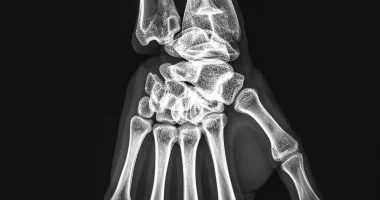
Diagnosis
When detecting the first signs of cubital canal syndrome, patients are advised to consult a neurologist for a thorough physical examination, conduct special tests, and obtain a referral for instrumental diagnostics. Among the numerous diagnostic tests that determine the damage to the ulnar nerve, the following are considered the most informative:
- When trying to extend the fingers in patients with cubital canal syndrome, the little finger, ring finger, and part of the middle finger do not fully extend;
- the patient is unable to “scratch” the table surface with the little finger while the palm is firmly pressed against it;
- the fingers of the hand cannot be brought in and out with the hand resting on the table surface.
To determine the causes of the development of pathological processes, the patient is prescribed additional instrumental studies:
- Electroneuromyography of the ulnar nerve detects lesions, and their localization has predictive value. It also allows the exclusion of other pathologies mimicking ulnar neuropathy. The evaluation of electroneuromyography results is carried out taking into account the existing clinical picture. Needle electromyography is additionally performed.
- Ultrasound of the ulnar nerve. Ultrasound ⎼ is an auxiliary diagnostic tool that determines the pinching site, assesses the condition of adjacent tissues, and detects cysts and other neoplasms.
- CT scan of the elbow joint. In cubital canal syndrome, computed tomography assesses the state of bone tissues, and the study is carried out to detect osteophytes, other posttraumatic consequences, and developing inflammatory processes.
- MRI of the elbow joint. It is used to visualize the causes of ulnar nerve impingement directly, determine the anatomical pattern of muscle denervation, and evaluate soft tissue conditions to characterize the lesion.
Computed tomography and magnetic resonance imaging of the elbow are not always indicated in patients with suspected cubital tunnel syndrome. These diagnostic procedures are helpful when there is reason to believe that there are concomitant elbow pathologies.
Treatment of cubital canal syndrome
Conservative therapy
Each patient is selected for an individual therapeutic course, considering the severity of the pathology. Conservative treatment is acceptable for mild and intermittent symptoms, but in severe cases, surgical intervention is recommended.
Conservative therapy is understood as a set of therapeutic measures, including:
- orthotics – preventing compression on the ulnar nerve by using special bandages that limit full flexion of the joint;
- drug therapy – prescription of non-steroidal anti-inflammatory drugs, drugs that normalize blood flow, B-group vitamins;
- physiotherapy, therapeutic massage.
Surgical treatment
Surgical intervention is resorted to when there is no expected effect from conservative treatment for three months or when the patient presents with a severe clinical picture. Surgical intervention to decompress the ulnar nerve is recognized as an effective treatment option with a high chance of success. Motor function of the extremities is usually restored after surgical treatment. Among the existing methods utilized are:
- simple decompression is an effective and safe surgical procedure with fewer postoperative complications for patients with cubital canal syndrome;
- Decompression with transposition is a surgical method of removing nerve compression while relocating the nerve to allow it to function normally;
- endoscopic decompression – is performed using special equipment that allows to perform operations in a less invasive way, eliminating significant blood loss and the risk of increased trauma;
- Medial epicondylectomy is a partial medial epicondyle excision technique to widen the cubital canal.
All these treatment options are available in more than 800 hospitals worldwide (https://doctor.global/results/diseases/guyons-canal-syndrome). For example, Guyon’s canal release can be performed in these countries at following approximate prices:
Turkey$1,400 in 32 clinics
Germany$5,185 in 40 clinics
China$6,385 in 6 clinics
Israel$7,172 in 16 clinics
United States$8,000 in 19 clinics.
Prognosis and prevention
Recovery is gradual due to the characteristic features of nerve tissue repair, so there is no need to wait for immediate results. It can be evaluated after several months, provided that all medical recommendations are followed. The speed of recovery is influenced by the duration of the previous compression. Treatment is successful in 70-80% of cases; recurrences are noted in about 25% of patients. It is believed that the effectiveness of surgical treatment is superior to conservative therapy.
As preventive measures, avoid traumatic situations, uncomfortable positions, increased joint stress, hypothermia, and excessive vibration. It is important to remember that the prognosis is much worse in neglected conditions. Untreated condition adversely affects the quality of life, leading to loss of work capacity and subsequent disability. Therefore, seeking medical help, early diagnosis, and therapy promptly prevents the onset of irreversible changes in the functions of the hand and fingers, as well as severe consequences.