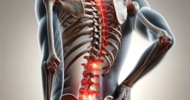
Hemifacial spasm
Define
Hemifacial spasm is a paroxysmal unilateral spastic contraction of facial muscles caused by various stimuli on the central intracranial portion of the facial nerve. It is diagnosed by peculiarities of clinical symptoms and characteristic electromyography patterns. Etiology is established by magnetic resonance imaging of the brain and cerebral vessels. Conservative (pharmacotherapy, botulinum toxin injection) and surgical (decompression of the nerve trunk, removal of formations compressing the nerve) techniques are used in treatment.
General information
The term hemifacial means “affecting half of the face”. Hemifacial spasm (HFS) was first described in 1975. Hemifacial spasm is diagnosed with an incidence of 0.8 cases per 100,000 population, predominantly in the 40-50 years of age. Women get the disease two times more often than men. Among persons of the mongoloid race, the incidence is higher, which is due to the peculiarities of the anatomy of the cranial box. Bilateral hemifacial spasm is observed in exceptional cases.
Causes of hemifacial spasm
The disease develops due to irritation of the root of the facial nerve coming out of the brain stem. Recent studies have shown that along with the typical localization of the irritation zone in the place of root exit, pathological effects on the nerve are possible throughout its central segment. The main etiologic factors causing adverse effects on the facial nerve are:
- Neurovascular conflict. It occurs at a special anatomical location of the nerve root and nearby arteries (lower cerebellar, basilar, vertebral arteries). Close contact of the nerve trunk with the arterial wall causes irritation due to vascular pulsation. Excessive excitation of nerve fibers causes hyperinnervation of the muscles of the corresponding half of the face.
- Aneurysms of cerebral vessels. Aneurysmal dilatations of the cerebral pontine vessels have a compressive effect on the facial nerve. As a result, the nerve fibers are over-irritated, provoking the occurrence of pathological impulsation to the facial musculature.
- Brain tumors. Tumors of the pontine-cerebellar angle can squeeze the facial root with their mass. As a result, excessive excitatory impulses flow along the nerve to the facial muscles, leading to spasms.
- Foci of demyelination. The spread of myelin destruction to the trunk of the facial nerve causes the transfer of excitation from one nerve fiber to another. Increased impulsation is formed, provoking muscle spasm.
- Area of lacunar infarction. In place of the ischemic area, there is the formation of a small cavity – a lacuna, which has an irritating effect on the facial root. Over-irritation of the nerve leads to impaired innervation of mimic muscles and their spastic state.
Classification
The division of facial hemispasm into two etiologic forms was formed historically due to insufficiently studied pathogenetic mechanisms of the disease. The introduction of neuroimaging diagnostic methods allowed us to identify the cause of the idiopathic form of the disease, but the division remained. According to the etiology, hemifacial spasm is classified into:
- Primary – caused by neurovascular conflict. Previously, primary hemispasm was considered idiopathic (no known cause).
- Secondary – caused by squeezing effect of volumetric mass (aneurysm, tumor, lacunae), demyelinating process.
The peculiarities of the clinical picture associated with the sequence of involvement in the spasm of the facial musculature formed the basis of clinical classification. Clinically distinguished:
- Typical HFS – spastic paroxysm begins with isolated contractions of the circular muscle of the eye. It is characteristic of the primary form.
- Atypical HFS – spasm occurs in the cheek muscles and spreads to the periorbital area and forehead. It occurs in thesecondary form.
Symptoms of hemifacial spasm
In the classic version, a spastic attack is started by contractions of the muscle around the eye. There are individual squeezes of the eye, the frequency of which increases. Gradually, the spastic contractions spread to all the muscles in one half of the face. Clenches become so frequent that the patient loses the ability to see with the affected eye. Atypical hemifacial paroxysm begins with the cheek muscles; then, the spasm spreads to the muscles of the lips, periorbital area, and forehead. Muscle contractions are painless and occur suddenly.
The hemifacial attack is provoked by psychoemotional tension and overwork. Hemifacial spasms can be observed in a sleeping patient, unlike other hyperkinesias that disappear in sleep. Babinski’s synkinesia symptom is considered pathognomonic: on the side of the lesion, eyelid closure is accompanied by eyebrow-raising, caused by simultaneous contraction of the circular muscle of the eye and the frontal forehead muscle.
The disease has a chronic course, accompanied by the gradual formation of moderate paresis of the facial musculature. In some cases, HFS is combined with the pathology of other cranial nerves. In 5% of patients, there is trigeminal neuralgia; in 15%, there is hearing loss of varying severity due to the lesion of the auditory root. Some patients complain of noise in the ear on the affected side, which occurs due to involvement in spasms of the muscle that moves the auditory ossicles.
Diagnosis
Hemifacial spasm is diagnosed by a neurologist based on clinical data: the nature of seizures, the presence of the synkinesia symptom, and the presence of paroxysms during sleep. The absence of other neurological symptoms is important. Patients with hearing impairment require consultation with an otolaryngologist. To confirm the diagnosis and establish the etiology of HFS, additional studies are carried out:
- Electromyography. Two types of myographic patterns characteristic of HFS have been described. A pathognomonic sign is the phenomenon of abnormal muscle response—at electrostimulation of one of the branches of the facial nerve, muscles innervated by its other branches contract.
- MRI of the brain allows diagnosing the cause of radicular compression: tumor, demyelination focus, vascular malformation, or lacuna. MRI data can also be used to judge the primary or secondary nature of HFS.
- Cerebral MR angiography is performed in addition to brain MRI to visualize neurovascular conflict. The latter’s detectability depends on the caliber of the conflicting vessel: for large vessels, it is 100%; for smaller vessels, it is 76%.
Treatment of hemifacial spasm
Conservative and neurosurgical techniques can be used. Conservative therapy is carried out for a long time, which leads to side effects. Neurosurgical treatment allows radically eliminating the cause of the disease but does not exclude recurrence.
Conservative techniques:
- Pharmacotherapy is carried out with antiepileptic agents and myorelaxants. Because the long-term results of drug therapy have not been sufficiently studied, some authors consider it doubtful.
- Botulinum toxin injection. Drug injections are made every 3-4 months subcutaneously and intramuscularly in the area of the affected muscle groups. The method is effective in 75% of patients and gives good results when starting therapy in the initial stages of the disease.
Neurosurgical interventions:
- Microvascular decompression is performed in case of neurovascular conflict. The operation consists of separating the nerve trunk and vessel with a Teflon protector in the place of their contact. The main postoperative complications are vestibulocochlear nerve dysfunction (2-3%) and facial paresis (3.5-4.8%), which is often transient.
- Surgical treatment of aneurysms. It is performed by clipping the neck of the aneurysm or its endovascular occlusion. It prevents rupture of the thinned vascular wall at the site of the bulge, leading to intracerebral hemorrhage.
- Removal of a cerebral tumor. It is performed using a microsurgical technique. The scope of surgery depends on the size and type of neoplasm and is adjusted intraoperatively based on the results of histologic examination.
All these treatment options are available in more than 600 hospitals worldwide (https://doctor.global/results/diseases/hemifacial-spasm). For example, Microvascular decompression (MVD) can be performed in these countries for following approximate prices:
Turkey $11.4 K in 28 clinics
China $24.4 K in 7 clinics
Germany $26.2 K in 39 clinics
Israel $29.4 K in 12 clinics
United States $40.1 K in 14 clinics.
Prognosis and prevention
Hemifacial spasms are characterized by a chronic course. There are known cases of spontaneous regression of symptomatology, amounting to 10%. According to various data, the recurrence of the primary form of the disease after microvascular decompression is up to 20%. The prognosis of the secondary form depends on the cause of the pathology. There is no specific prevention. Preventing vascular changes contributing to neurovascular conflict includes measures to prevent hypertension and atherosclerosis.