Osteochondrosis
What is osteochondrosis?
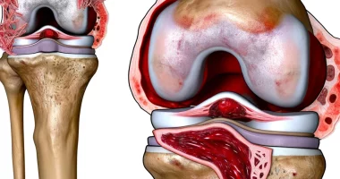
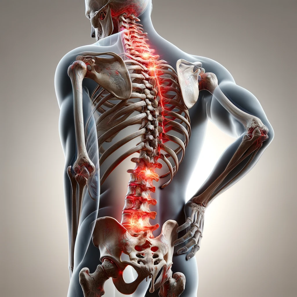
Osteochondrosis is a disease of the musculoskeletal system, manifested by thinning of bones and cartilage of joints. Most often, patients are diagnosed with osteochondrosis of the spine, in which the intervertebral discs are destroyed in the cervical, lumbar, or thoracic region. Intervertebral cartilage is necessary to cushion movements and reduce the load on bone tissue, so the destruction of discs leads to gradual damage to the vertebrae and deformation of the spine. Osteochondrosis can be a complication of trauma, long-term violation of posture, or other pathological conditions. Treatment of the disease involves manual therapy, physiotherapy, and surgical interventions for significant spinal deformities.
About the disease
Osteochondrosis is one of the most common diseases of the musculoskeletal system in the elderly. In adolescents and young people, pathology is diagnosed less often. To the characteristic symptoms of the disease, doctors include severe pain in the area of the affected joint and impaired mobility. Without treatment, osteochondrosis can be complicated by compression of the spinal cord and violation of the functions of internal organs.
Types
The most common classification of osteochondrosis depends on the localization of changes. The following types are distinguished:
- cervical;
- thoracic;
- lumbar.
The severity of pathological changes allows us to distinguish three stages of disease development:
- Stage 1: there is flattening and slight bulging of the intervertebral disc;
- Stage 2: cracks appear in the fibrous ring surrounding the disc, and the affected spinal segment loses stability;
- Stage 3: the fibrous ring is torn and a herniated disc is formed.
Symptoms of osteochondrosis.
The manifestations of the disease depend on the area of destruction of the articular discs. The main symptom is aching or sharp pain that increases during movements. Because of this, patients often assume a forced posture. With osteochondrosis of the cervical spine, it is difficult for the patient to look around, and there are severe headaches. If the disease is complicated by compression of peripheral nerves and spinal cord, there are symptoms of impaired movement and disorders of the functions of internal organs.
Common signs of osteochondrosis include:
- moderate dull pain during the day, increasing during movements;
- spread of pain to the head, shoulder, pelvis, and lower extremities;
- constant strain on the muscles associated with the affected intervertebral joint;
- decreased performance;
- sleep disturbance;
- changes in posture and clumsy gait.
The disease can progress over several years. The early stages of cartilage damage do not show any symptoms, but gradually, there is constant soreness.
Osteochondrosis of the cervical spine
The main symptom is pain caused by lateral compression of the spinal cord roots by bony growths. Unusual sensations and muscle dysfunction often accompany pain. The volume of movements in the neck is disturbed; they become limited and accompanied by pain and crunching.
Clinical picture of osteochondrosis of the cervical spine:
- constant or fulgurant (in the form of shooting pains) pain in the neck; the pain is intense, intensifies after sleep and with sharp turns of the neck;
- tension in the neck muscles, more in the anterior group;
- smooth cervical lordosis, forced position of the head (often tilted toward the side of the disc lesion);
- compression of the vertebral sympathetic plexus and impaired patency of the arteries in the spinal canal;
- Circulatory disorders in the cerebellum, brain stem, and occipital lobes of the brain: characterized by constant headaches; sometimes the pain is accompanied by disorders in the form of nausea or vomiting, sensations of noise in the head, ringing in the ears, synchronous with the pulse, visual disturbances in the form of flickering, pain in the eye, may feel a foreign body in the throat.
Osteochondrosis of the thoracic spine
Osteochondrosis of the thoracic spine is characterized by pain in the interscapular region. Often, the pain in the chest becomes shingles and is felt along the course of the intercostal sutures. Superficial and deep sensitivity may decrease.
Angina pectoris, pain in the liver region, and gastrointestinal dysfunction manifest visceral (internal) disorders. Sometimes, there are disorders of urination and sexual function.
Osteochondrosis of the lumbar region
The most common type of osteochondrosis is lumbar osteochondrosis. There is pain syndrome in the lumbosacral region with pain spreading to the legs (lubmoischialgia) or with localization only in the leg (ischialgia).
Pain, predominantly dull and aching, increases with sudden movements and prolonged forced posture and decreases with the horizontal position of the torso. Possible atrophy and paresis (loss of sensation), decreased reflexes.
Dry and flaky skin, sweating disorders, and skin lividity manifest vegetative disorders. Static disorders manifest by flattening the lumbar lordosis as an adaptive reaction to reduce the volume of spinal motion.
Causes of osteochondrosis
Usually, intervertebral cartilage is continuously supplied with blood and receives sufficient nutrients from the blood to renew cells and maintain its structure. As we age, degenerative changes may occur in the tissue, characterized by insufficient cell renewal. Violation of blood flow occurs when arteries are compressed against the background of constant curvature of the spine or trauma. Also, osteochondrosis can be the result of excessive physical exertion.
Among the most common causes of the disease are:
- The gradual destruction of intervertebral cartilage due to poor posture, weight lifting, and movement patterns, prolonged wearing of flat-soled shoes;
- craniovertebral anomalies – a violation of the mutual arrangement of the structures of the cervical spine and skull; this often results in compression of blood vessels and impaired blood supply to the tissues;
- metabolic disorder accompanied by insufficient formation of hydrophilic components of cartilage;
- An inflammatory or autoimmune cartilage lesion in which the body’s immune system attacks healthy tissue.
Clarifying the cause of osteochondrosis is essential for selecting the proper treatment.
Diagnosis of osteochondrosis
When symptoms of osteochondrosis appear, it is necessary to make an appointment with a neurologist. Doctors usually ask the patient about complaints and study the medical history to detect risk factors for the disease. Then a general examination is carried out to identify external signs of osteochondrosis and exclude symptoms of spinal cord dysfunction. To confirm the diagnosis, the neurologist prescribes special examinations.
- Radiography of the spine. The neurologist prescribes this study to obtain an image of the affected spinal segment in two projections. Before the procedure, the patient is asked to undress to the waist and stand in front of the apparatus. The resulting images allow the doctor to detect herniation or other pathology, as well as to determine the degree of damage to the vertebrae and intervertebral disc;
- Magnetic resonance imaging is a more informative study that allows the neurologist to examine layer-by-layer images of the spine in different projections. The patient is asked to remove all metal jewelry and lie on the tomography table during the study. Diagnosis takes from 15 minutes to an hour. This study is a painless and reliable method of diagnosing osteochondrosis. The MRI results make it possible to immediately exclude other diseases with similar symptoms, such as tuberculous bone lesions or malignant tumors. If MRI is contraindicated, myelography is indicated.
- Examination of the subarachnoid space of the spinal cord (myelography). A neurologist prescribes this procedure to exclude syringomyelia, oncology, and other pathological conditions of the brain membranes. The method of obtaining an image does not differ from the usual radiography, but before the study, the doctor injects a contrast agent into the subarachnoid space. Before puncture, the doctor treats the skin with antiseptic and anesthetizes the tissue. The needle is inserted into the intervertebral space of the lumbar spine.
Treatment
Conservative treatment of osteochondrosis
Treatment is carried out after consultation with a neurologist and orthopedist. In case of instability of the affected cervical spine segment, a Shantz collar is applied. A pre-made orthopedic collar is applied to the neck and strengthened with bandages. Wearing a collar is necessary to ease the load on the affected spine and prevent the development of complications. The patient needs rest in severe osteochondrosis of the lumbar or thoracic spine.
Depending on the specific clinical situation, doctors use different therapeutic techniques.
- Administration of non-steroidal anti-inflammatory drugs for pain relief. Strict control of analgesic dosage and liver monitoring is required for prolonged use of analgesics.
- Traction therapy is a method of stretching the spine to widen the intervertebral gap to eliminate compression of blood vessels and nerves. Before the procedure, a massage is performed to relax the muscles. Stretching is performed on a special table or in a bathtub. The device is fixed on the patient’s body in the desired position and gently stretches the spine. This is a very effective and safe procedure for treating osteochondrosis.
- Use of anticonvulsant medications for complications of osteochondrosis.
- Use of myorelaxants. These medications relax the muscles associated with the affected spine and relieve pain;
- Use of corticosteroids for nerve compression. These drugs relieve the inflammatory process in the tissues.
- Physiotherapy involves therapeutic massage, special exercises, and warming.
A neurologist selects an individual therapy regimen for osteochondrosis based on the patient’s condition. Strict medical control of the treatment course allows for the best prognosis.
Surgical treatment of osteochondrosis
If the diagnostic results reveal an intervertebral hernia or severe compression of the spinal canal, the patient is prescribed a consultation with a neurosurgeon. The doctor assesses the severity of the detected pathology and, if necessary, performs surgical intervention.
- Partial or complete removal of the intervertebral disc. The operation is performed under anesthesia. After applying anesthesia, the neurosurgeon treats the operating field with antiseptic and cuts the skin in a predetermined place. After sliding the muscle layer with a special instrument, the doctor removes the affected intervertebral disc. If necessary, removal of the vertebral arch is also performed. Plasticization of the vertebral segment with transplantation of artificial materials is performed. This effective surgery is suitable for the treatment of intervertebral herniation and for alleviating the symptoms of osteochondrosis;
- Surgery for narrowing of the spinal canal. This intervention eliminates the compression of nerves and spinal cord. After applying anesthesia, the doctor treats the operating field with antiseptic and makes a wide skin incision. The neurosurgeon reaches the intervertebral joint and performs manipulation by pushing away the muscle layer. The intervention involves the removal of the vertebral arch or the entire joint. X-ray and microsurgical control allow the operation to be performed with minimal risks.
All these treatment options are available in more than 750 hospitals worldwide (https://doctor.global/results/diseases/osteochondrosis). For example, cervical laminectomy is performed in 31 clinics across Turkey for an approximate price of $6.7 K (https://doctor.global/results/asia/turkey/all-cities/all-specializations/procedures/cervical-laminectomy).
Prevention
Prevention methods include:
- Wearing comfortable shoes with cushioned soles;
- moderate exercise and maintaining good posture;
- regular checkups with an orthopedist for spinal curvature.
Rehabilitation
The recovery duration after surgical treatment depends on the type and complexity of the surgery. As a rule, in the first few days, the patient follows bed rest and then gradually increases physical activity under the supervision of doctors. In the first stages, it is necessary to wear a special corset, limiting the load on the spine. As the condition stabilizes, the patient is referred to physical therapy, therapeutic physical training, etc.