
Radial shaft fracture
What’s that?
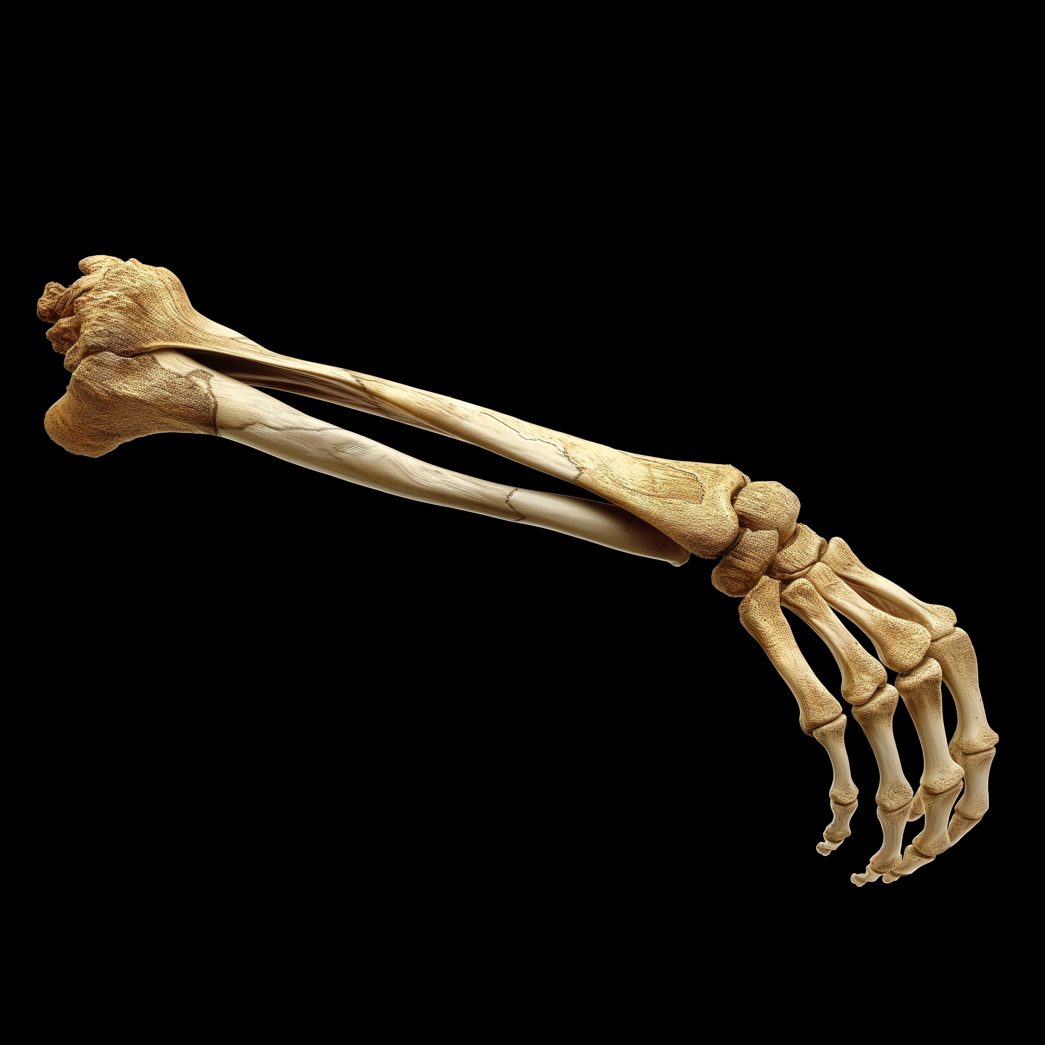
A radial shaft fracture, or fracture of the radius, is a frequently encountered injury of the hand. This condition is characterized by a violation of the integrity of bone tissue under the influence of mechanical load of varying intensity.
About the disease
The radius is most often broken in the so-called typical place. This area is located 2-2.5 cm above the articular line of the wrist joint, and the ulnar styloid process is often injured as well. This injury is categorized as complex and presents particular difficulties for treatment.
Fractures of the radial head are intra-articular, often accompanied by crushing and formation of many fragments. This results in limitation of flexion, extension, forearm rotation, and long-term disability. Fractures of the radial head are often treated surgically.
Fractures of the body of the radius are rare. They are usually combined with trauma to the ulna. Such conditions are associated with a direct impact on the forearm bones.
Radial fractures are manifested by classic signs – pain, swelling of tissues, hemorrhages in soft tissues, unnatural forearm mobility, and bone crunch. In the presence of displacement of broken segments, the shape of the arm is disturbed. The final diagnosis is based on visualization of the damage zone using X-ray scanning.
The nature of treatment depends on the complexity of the injury. A conservative approach is practical in simple fractures, while complex fractures require surgical intervention.
Types
Taking into account the multiplicity of lesions, they are distinguished:
- Isolated injuries where only the radius is broken;
- Combined injuries, when radius fractures can be combined with bone trauma of other localization (e.g., damage to bones of the leg, spine, ribs, etc. in car accidents).
According to localization, the following types of traumatic radius injury are distinguished:
- An injury in a typical location in which the end of the bone (epimetaphysis) closest to the wrist joint (distal process) is injured;
- Diaphyseal injury of the radius;
- damage in the area of the head or neck of the radius, i.e., those areas closer to the elbow joint (proximal segment).
Depending on the condition of the skin over the site of injury, fractures are of 2 classes:
- open, when not only the bone but also the skin is damaged (having an open wound increases the risk of infectious complications);
- closed, in which the skin is intact (only the integrity of the bone is compromised).
According to the location of the bone fragments are distinguished:
- displaced injuries – the fragments are located at different angles to each other, which is not physiologic for this bone (also possible puncture fractures, when one fragment goes over the other, which reduces the length of the limb);
- injuries without displacement – broken segments retain the typical direction of the bone.
Intra-articular fractures are a separate category in which the fracture line runs inside the joint cavity. Both the wrist and elbow joints can be damaged.
Fractures can be compression fractures (an indentation zone is formed), impression fractures (broken fragments are pushed inward), spiral fractures, etc.
Symptoms of a radius fracture
Symptoms of a radius fracture may include the following:
- intense pain in the injured area;
- swelling and lividity of the soft tissue;
- Increased pain when palpating the pathologic area, attempted movements in the joint;
- bone crunch;
- abnormal mobility;
- impaired functional status of the hand.
These symptoms are most significant in the injured area. Thus, if the integrity of the head or neck is violated – this is the area of the elbow joint, with a distal fracture – the area of the wrist joint. If the body of the radius is injured, then the middle third of the forearm swells and hurts.
Reasons
Fractures of the radius can result from a household (fall) or sports injury, transport accident, or brunt.
- Radial fractures in the typical zone are one of the most frequent types of bone injuries. The mechanism of such an injury is associated with a fall on the arm extended at the elbow. If the hand was in a state of extension, an extension (extensional) fracture was formed; if in a state of flexion, a flexion (flexion) fracture was formed. Typical fracture of the radius is often found in postmenopausal women, which is associated with systemic osteoporosis. Therefore, it is essential to carry out timely prevention of bone fragility.
Diagnosis
The preliminary diagnosis of radius fracture is established based on the data of objective examination in combination with the indication of the fact of injury. The final diagnosis involves radiography in at least two projections. X-ray allows you to clearly visualize the line of injury, assess the nature of the injury, and develop a program for further patient treatment.
In complex clinical cases, computerized or magnetic resonance imaging is performed. If menopausal osteoporosis is suspected, densitometry, which determines the strength of bone tissue, is indicated, as well as laboratory tests reflecting the level of calcium and vitamin D in the blood.
Treatment of radius fracture
Conservative or surgical methods can be used to treat radius fractures. The treatment program choice is determined by the type of injury, the patient’s age, professional activity, and concomitant pathology.
Conservative treatment
Conservative treatment of fractures of the distal and proximal processes is quite long, with an average of 6 to 10 weeks (immobilization in the repositioning position, plaster cast after the limb is removed from the repositioning position and rehabilitation treatment). The period of rehabilitation increases depending on the duration of fracture fusion, duration of rehabilitation treatment, and trophic disorders. Therefore, physically active patients with such injuries are often treated surgically.
Conservative management of fractures is often used when the radial bone body is injured (with injuries of the epiphysis and metaepiphysis, the issue is solved individually). In the presence of displacement of bone fragments, their manual repositioning under anesthesia is first performed. After that, the effectiveness of repositioning is evaluated using radiography, and if the result is satisfactory, a circular plaster cast is applied. In the absence of displacement, plaster immobilization is performed immediately. The average healing time of diaphyseal fractures of the radius is 3-4 weeks.
Surgical treatment
Surgery for fracture of the radius in a typical location is superior to conservative treatment in terms of efficacy and other indicators. Among surgical methods, open repositioning with plate fixation is favored. It makes it possible to restore the anatomical integrity of the bone and function in the adjacent joints in the shortest possible time without waiting for complete fracture fusion and to avoid possible complications (trophic disorders, contractures of the wrist joint), the probability of which increases with increasing immobilization time.
When treating a fracture of the radial head, the surgeon can perform a juxtaposition of the fragments and their periosteal fixation with special plates and screws.
All these treatment options are available in more than 800 hospitals worldwide (https://doctor.global/results/diseases/radial-shaft-fracture). For example, forearm fracture surgery is performed in 40 clinics across Germany for an approximate price of $7.3 K (https://doctor.global/results/europe/germany/all-cities/all-specializations/procedures/forearm-fracture-surgery).
Prevention
The prevention of radial fracture involves preventing injuries. It is essential to take safety precautions during icy conditions, when driving, and to use protective equipment when participating in injury-prone sports.
Menopausal women are recommended to determine the 10-year fracture risk by densitometry using a special algorithm. If the risk is high, menopausal hormone therapy is considered in conjunction with an obstetrician-gynecologist. At the same time, if a woman has premature menopause (before the age of 45 years), even without calculating the risks, preventive prevention of osteoporosis with natural estrogens of systemic action is carried out. It makes it possible to prevent pathologic fractures due to low-energy exposure.
Rehabilitation after radius fracture
The rehabilitation program for radius fractures consists of the following areas:
- physical therapy exercises aimed at developing the joint and strengthening muscles;
- Physical treatments that help improve blood circulation and stimulate bone regeneration;
- A revitalizing massage that tones muscles and relieves swelling.
Rehabilitation after a radius fracture begins almost immediately after surgery, as plaster immobilization is unnecessary. Physical therapy is recommended from the second day after surgery. On the 5th to 7th day after surgery, the wrist or elbow joint movement volume is close to complete. The total rehabilitation period is generally 3-4 weeks.
If the bones were fused by creating immobility with a cast, active rehabilitation begins only after the bandage is removed.
