Spinal fracture
What’s that?
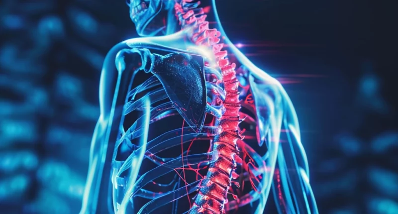
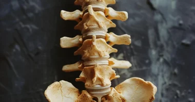
A spinal fracture is a bone injury that involves one or more vertebrae (bodies or processes).
About the disease
Vertebral fractures can be the result of traumatic injury or osteoporosis, which is characterized by decreased mineralization of bone tissue, making it more fragile. Vertebral fractures are manifested by localized pain. Taking into account the characteristics of the damage can also violate sensory and motor innervation and upset the work of the pelvic organs. In severe cases, paresis or paralysis develops.
Diagnosis of spinal fracture is based on the results of objective examination and X-ray scans with direct and lateral scans. In some situations, computed tomography (CT) or magnetic resonance imaging (MRI) is indicated.
Considering vertebral injury’s clinical and radiologic features, conservative treatment or surgical intervention may be indicated. Surgery allows to restore the anatomy of the damaged vertebra and cope with signs of spinal cord compression.
Types of spinal fractures
According to the etiologic cause, the following types of spinal fractures are distinguished:
- traumatic – caused by high-energy impact from the outside;
- pathological – developed against the background of primary changes in the structure of bone tissue (for example, against the background of osteoporosis, tumor, or infectious process).
Vertebral fractures are categorized into two types based on their degree of danger:
- uncomplicated – the condition is characterized only by a violation of the integrity of the bone base;
- complicated – the fracture may be accompanied by displacement of bone segments with subsequent compression of the spinal cord or nerve roots.
According to localization, vertebral fractures are divided into the following types:
- cervical trauma;
- thoracic trauma;
- lumbar trauma;
- trauma to the sacral or coccygeal segment.
According to the condition of the skin over the site of traumatic injury, vertebral fractures can be open (there is a soft tissue wound) or closed (the skin is intact).
By nature of injury, vertebral fractures can be of 3 types:
- Type A – compression fractures in which compression develops in the anterior segment of the vertebra (may be comminuted fractures);
- Type B – distraction fractures in which there is a separation of the bone fragments;
- Type C – rotational fractures with or without displacement of bone fragments.
Symptoms
The main clinical signs of a fracture may include:
- painful sensations in the area of traumatic impact;
- painful sensations when turning the head, lifting or lowering the limbs;
- disruption of pain or tactile innervation, which is determined by the level of spinal cord injury;
- impaired mobility of the spine (flexion is usually the first to suffer);
- impaired motor activity of the arms or legs;
- symptoms of spinal shock.
If the vertebrae in the cervical vertebral column are injured, the victim reflexively tries to stretch the neck to reduce the pressure on the injured vertebrae. In some cases, the person supports their head with their arms. Damage to the thoracic or lumbar section is accompanied by an unnatural straightening of posture, with an attempt to sit down accompanied by intense pain (a person reflexively holds the body with his hands to shift the load on the sciatic tubercles). In some cases, lumbar trauma leads to the appearance of pseudoperitonitis – symptoms of peritoneal irritation.
Reasons
In the total structure of fractures in the human body, approximately 1.5-2% are vertebral fractures. The main factors oftraumatic fractures can be:
- transportation accidents;
- falling from a height;
- sports trauma.
Vertebral integrity can be compromised by direct or indirect impact. The direction of the fracture line can be different, depending on the injury mechanism. Thus, there is a distinction between flexion or extension fracture, rotational and compression fracture (in the latter case, the load is exerted along the axis, and the bone segments seem to be pressed into each other).
Fractures of the spinous processes are caused by both direct (impact on the spinous process) and indirect (overextension or sudden flexion of the spine) mechanisms of injury. Several spinous processes may be fractured at once. Above the area of injury, swelling of tissues is detected, and palpation determines severe pain. Palpation of the line of the spinous processes allows the change in the distance between them to be determined; the damaged process has unnatural mobility and deviates from the median line to the side.
Causes of vertebral fractures in older people are usually due to age-related osteoporosis.
Diagnosis of a spinal fracture
A spinal fracture diagnosis program may include the following methods:
- an objective examination by a trauma surgeon or neurosurgeon;
- radiography in at least two projections;
- neurological examination to rule out damage to nerve tissue (spinal cord or roots);
- computed tomography or CT myelography.
Treatment of spinal fracture
The characteristics of the injury determine the treatment of a spinal fracture. A-type fractures are treated primarily with conservative care, while B-type fractures require stabilization of the spinal column and, in some cases, spinal decompression. Type C injury always requires decompression and rigid fixation of the vertebral column.
Conservative treatment
First aid and pre-hospital care:
- is performed only when the victim is horizontal;
- fixation on a rigid surface;
- additional fixation of the neck area with a Shantz collar;
- attempts to sit down and any spinal column movement are strictly forbidden.
If a regular (non-rigid) stretcher is used for transportation, the person lies on the stomach. A bolster is placed under the shoulder area to raise the head.
Subsequent conservative care involves traction and rigid fixation if necessary.
Surgical treatment
Surgery for a spinal fracture has several purposes:
- to prevent compression of spinal structures;
- fixation of bone fragments to accelerate healing;
- ensuring early rehabilitation;
- prevention of delayed curvature of the spinal column;
- pain alleviation.
The nature of the injury determines the specifics of surgical intervention. Surgery can be performed for both fresh and long-standing injuries.
All these treatment options are available in more than 800 hospitals worldwide (https://doctor.global/results/diseases/spinal-fracture). For example, Instrumented spine stabilization is performed in 31 clinics across Turkey for an approximate price of $13.6 K (https://doctor.global/results/asia/turkey/all-cities/all-specializations/procedures/instrumented-spine-stabilization).
Prevention
Preventing vertebral injuries focuses on following safety rules while driving, sports activities, and household chores.
Rehabilitation after a spinal fracture
Rehabilitation after a spinal fracture usually begins the day after surgery:
- on the second day of the postoperative period, it is allowed to get up and walk;
- use a semi-rigid removable brace for verticalization;
- wearing the corset is usually recommended for three months;
- refrain from lifting heavy objects and working in an inclined position for three months;
- avoid excessive exposure of the operated patient to cold or heat for three months;
- after surgery, do simple physical exercises selected by a rehabilitation specialist;
- include physical therapy and massage as part of the recovery program.


