
Pleural effusion
What is it?
Hydrothorax, or pleural effusion – is an accumulation of fluid (transudate) in the lung area not associated with inflammatory processes. Hydrothorax is not an independent disease. It is a symptom accompanying several diseases. As it progresses, it provokes a breakdown in respiratory function and changes the shape of the lungs. You should consult a specialist to prevent complications and identify the underlying disease.
Disease
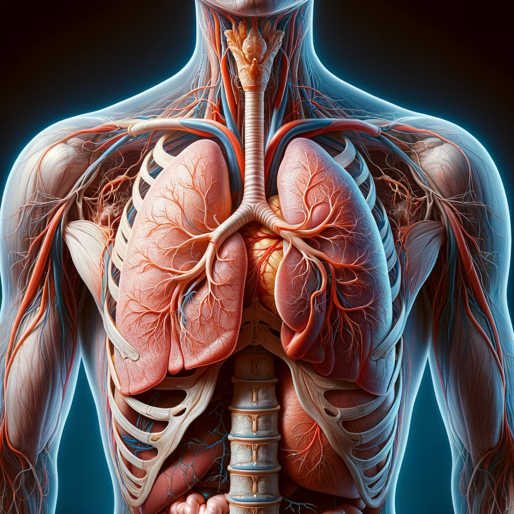
Protecting the lungs inside the body is presented by a double-layered sheath – pleura. It consists of an outer and an inner layer. There is a narrow space between them – the pleural cavity. Usually, pleura produces a certain amount of fluid. Its total volume is about 20 ml.
This fluid is necessary to keep the lungs from rubbing against nearby structures when breathing. An increase in its amount indicates the development of pathologies. Most often, they are related to the cardiac system.
Hydrothorax should be distinguished from pleuritis – an inflammation of the pleura that occurs when the layers of the pleura are inflamed.
Classification
Effusion can be left-, right- and both-sided. Right-sided hydrothorax is an accumulation of fluid in the right pleural space. This type is most often diagnosed. Left-sided hydrothorax is an accumulation of fluid in the pleural space of the left lung.
Bilateral hydrothorax is affection of both lungs in this process. Fluid accumulation can be symmetrical (at one level) or asymmetrical (at different levels).
Depending on the extent of effusion between the pleural layers, encysted, partial, and total types are defined. Encysted hydrothorax occurs when fluid accumulates in space, confined by adhesions. It can form in any part of the shell. Total hydrothorax covers the entire area of the lung, the partial – only one part.
Hydrothorax types depending on volume:
- Small: fluid accumulates in the outer lower part of the pleura – in costodiaphragmatic sinus. For bilateral small hydrothorax, both corners of the lungs will be symmetrically darkened on X-ray.
- Medium: the liquid takes up the volume up to the middle of the scapula. Accumulates up to 1.5 liters.
- Large: fluid rises above the level of the scapula.
According to the severity of symptoms, hydrothorax can be acute, subacute, or chronic.
Symptoms
Symptoms depend on the amount of fluid, its localization, and the disease that provokes the effusion. The main sign is progressive shortness of breath. The lungs compressed by the effusion cannot fully expand; thus, they become airless. Initially, the symptom only appears during exercise, when the body requires an increased amount of oxygen. Further, shortness of breath begins to trouble and at rest.
Other symptoms of hydrothorax include:
- discomfort in the chest: the condition is relieved by lying on your side;
- pain in the sternum during inhalation;
- increased breathlessness when lying on the back; relief can be achieved in a sitting or semi-sitting position;
- intense cough;
- pallor of the skin, nasolabial triangle cyanosis.
There may be a heart rhythm disorder and abdominal pain because fluid can accumulate in the chest, pericardium, and abdominal cavity.
Symptoms increase depending on the amount of fluid accumulation. Once infected, the temperature rises. In some cases, the syndrome is asymptomatic.
Causes
The leading causes of pulmonary hydrothorax include the following conditions.
- Congestive heart failure. Due to impairment in the heart muscle contraction, blood is pumped slowly through the pulmonary arteries, pressure increases, and fluid leeks through the walls into the pleural cavity. In 80% of patients with chronic heart failure, congestive fluid accumulation occurs on both sides.
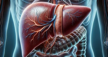
- Liver cirrhosis, kidney failure. Damage to the functional tissues of these organs leads to a breakdown in protein metabolism, which alters the amount of fluid filtering through the blood vessel walls. As a result, leaks appear not only in the lungs but also in the abdominal cavity. In liver damage, in 85% of cases, a moderate to small hydrothorax occurs on one of the sides.
- Pulmonary thrombus. It leads to a breach in the permeability of the blood vessel walls.
- Tumors compressing the tissues and blood vessels adjacent to the lungs.
- Autoimmune systemic diseases of connective tissue.
- An anatomical defect – a pathological communication between the pleura and the abdominal cavity.
Diseases that can cause hydrothorax: rheumatoid arthritis, rheumatism, pancreatitis, pulmonary or heart infarctions.
Diagnostics
The primary diagnosis is performed by a general practitioner or pulmonologist. Physical examination reveals thoracic asymmetry and limited lung mobility at the side of effusion. The doctor listens for natural breathing and taps the surface of the chest to determine the localization of the fluid.
The algorithm for further investigations includes several procedures.
- Ultrasound of the lungs. It allows you to measure the amount of fluid and mark the site of its puncture.
- Chest X-ray. Signs of fluid accumulation include uniform light spots (darkening) with a distinctly bordered descending ridge. The shape of this spots change while breathing.
- CT is the most informative way to determine the cause of the effusion.
- Diagnostic thoracocentesis – puncturing the pleural cavity at the same time as taking a fluid sample for examination.
Based on the results of the primary diagnosis, the doctor may order an ultrasound of the kidneys, abdominal organs, thyroid gland, ECG, and general and biochemical blood tests. An endocrinologist, hepatologist, cardiologist, or nephrologist is involved in the diagnosis if necessary.
Treatment
Conservative and surgical methods are used for the treatment of hydrothorax. The choice depends on the symptoms’ severity and underlying disease type. In most cases, signs of shortness of breath disappear after the replacement provoked by the pathology.
Conservative treatment
Conservative methods are mainly used to treat benign small hydrothorax. The patient may be prescribed:
- potassium-sparing diuretics to remove excess fluid;
- antibiotics for the infection and after the puncture;
- hormones to normalize the endocrine system;
- painkillers and antihypertensive drugs.
With effusion, significant help can be provided by correcting the diet. It is recommended to avoid fatty, acidic, spicy foods, legumes, and baked goods. Salt intake should be minimized.
Surgical treatment
To relieve symptoms and free up the lung, the puncture is performed under local anesthetic, and excess fluid is pumped out. Indications: medium to large volume of fluid, shortness of breath. In the case of recurrent accumulation, a drainage system is installed to ensure the continuous discharge of fluid.
If both methods are ineffective, pleurodesis is performed. After pumping the fluid out, a sclerosing (gluing) agent is introduced into the cavity, which solders pleural layers together.
Video-assisted thoracoscopic surgery (VATS) is used in severe cases, during which defects in the diaphragm and other causes of effusion are eliminated.
All these surgical procedures are performed in more than 530 hospitals worldwide (https://doctor.global/results/diseases/pleural-effusion). For example, thoracentesis can be done in 27 clinics across Turkey with an average price of $721(https://doctor.global/results/asia/turkey/all-cities/all-specializations/procedures/thoracentesis)
Prevention
To prevent hydrothorax, it is necessary to cure heart, kidney, and liver diseases. At the first sign of pain in the back of the sternum or peritoneum, you should consult your doctor. Only a doctor can tell what is happening to the patient, whether it is hydrothorax, what type it belongs to (large, small, right-sided, left-sided, bilateral), and how to treat it.
Even a diagnosed effusion, even a small amount, requires specialist supervision. It is essential to follow the diet and other doctor recommendations.
Rehabilitation services
After performing pleurodesis, drainage is installed for 2-4 days to drain the remaining liquid. During this time, the patient remains in the hospital. He is prescribed antibiotics and painkillers. After four days, a control X-ray is taken. Until complete healing, exercise, and swimming are contraindicated.