Introduction
Impingement syndrome of the ankle joint is a pathological condition in which its denser structures compress the joint’s soft tissues. At first, such a disorder can be characterized by discomfort in the damaged joint area, which can later develop into a pain syndrome. Lack of treatment for the disease leads to a decrease in the function of the ankle joint and damage to the intra-articular structures: cartilage, ligaments, and joint capsule.
If you suspect impingement syndrome, we recommend that you consult a doctor. Complications can be prevented by detecting initial changes in the joint and periarticular area in the early stages of the disease.
Impingement syndrome of the ankle joint is a pathologic condition resulting from a conflict in motion of the articular surfaces of the tibia and talus and the tissues surrounding the ankle joint.
It is one of the most common lesions of the ankle joint, consisting of impingement of bone or soft tissue structures between the joint’s articular surfaces. This condition can be caused by adhesions, osteophytes, osteochondral lesions of the talus bone, and many other causes. In patients, it manifests itself, in addition to pain syndrome, with a limited range of motion in the ankle joint.
This syndrome occurs when the space between the articular surfaces of the bones that make up the ankle joint is reduced. It occurs as a result of congenital predisposition (a certain shape of the talus and the articular surface of the tibia), as a result of trauma (post-traumatic displacement of bone fragments, rupture of the ligamentous apparatus), or a chronic inflammatory process in the ankle joint, resulting in overgrowth of the synovial membrane, which is pinched during movements in the joint. In addition, constant minor trauma causes ankle impingement syndrome. Patients involved in active sports are in the risk group.
Causes
Impingement syndrome of the ankle joint has many causes. Sprains often cause blockages of the forefoot. Also, ankle sprains can cause thickening of the outer ligaments. When the foot is lifted repeatedly, the ligaments can become pinched. Such an injury leads to limited mobility of the anterior ankle joint. In addition, this pathology can provoke irritation of the joint capsule. One of the painful consequences of impingement syndrome is synovitis (inflammation of the synovial membrane) of the entire ankle joint.
Due to instability, bone spurs (osteophytes) sometimes appear on the anterior edge of the tibia. Therefore, removing bony structures in this ankle segment in case of impingement is a joint-preserving manipulation.
Symptoms
Asymptomatic manifestation of impingement syndrome is a very rare phenomenon. In the presence of pathology, the patient feels restricted and stiff joint movements, as well as a pronounced pain syndrome.
Symptoms of the disease include:
- A change in the patient’s gait,
- Limitation of joint mobility,
- Decreased muscle tone,
- Swelling at the site of the injury,
- Painful sensations that increase with movement,
- In the neglected stages of the disease, joint deformation is manifested.
In addition to the symptoms mentioned above, the attending physician must assess the family history to identify the likelihood of hereditary joint pathologies.
Risk factors
Regardless of the site of injury, the following factors predispose to impingement syndrome_:_
- Diseases of the endocrine system,
- Diabetes mellitus,
- Hormonal disorders,
- Gout,
- Intense training and professional sports,
- Engaging in heavy physical labor.
Types of ankle impingement syndrome
Depending on the location of the lesion, anterior and posterior impingement are distinguished. Impingement of the ankle joint develops due to impaired mobility of the articular surface of the talus and tibia. The tissues surrounding the joint are also involved in the process. There is impingement of bone and soft tissue between the articular heads. Anterior manifests itself with pain in the anterior part of the joint and occurs due to traumatization of the joint. Soccer players are most often affected.
Posterior, respectively, is manifested by pain in the back and occurs in those people who walk on their toes or tiptoes for long periods of time, such as ballet dancers.
Diagnosis
At the initial stages of diagnosis, anamnesis is collected, and the injured limb is examined. Subsequently, a number of laboratory and instrumental examinations are performed to more accurately diagnose and confirm the presence of impingement syndrome.
Obtaining the most accurate data about the disease is achieved with:
- X-ray;
- CT;
- MRI.
Specialists additionally perform specific impingement tests to clarify the severity of the disease and determine the level of movement restriction.
Degrees of disease
Impingement syndrome is categorized into three stages depending on the severity of the disease:
- Stage 1. The integrity of the tendon is intact. Bleeding occurs, and soft tissue edema develops.
- Stage 2. On medical examination, foci of fibrosis begin to be fixed in the tendon structure, and often, tears occur. Inflammation becomes chronic.
- Stage 3. Bony growths – osteophytes – form in the damaged area. Because of this, degenerative changes occur in the surrounding soft tissues. Even if the patient provides minimal load on the limb, there is a high risk of tendon rupture.
Treatment
Specialists treat impingement syndrome using arthroscopic low-traumatic intervention – resection of osteophytes. During the operation, the surgeon makes several small incisions no larger than 5 mm. Penetrating them with an arthroscope equipped with a micro-camera, the doctor can examine in detail the internal state of the joint and immediately decompress the nerve (intra-articular) structures. Ankle impingement usually involves the removal of a bone spur at the edge of the tibia using an arthroscopic method. The osteophytes are removed using a special cutter. During arthroscopy, the orthopedic surgeon inserts special instruments into the joint through small incisions in the skin on both sides of the ankle. One instrument is an arthroscopic camera that shows an image of the surgical field on a large video screen. Instruments such as forceps, graspers, and medical aspirators are inserted into the surgical area through another tube. Ankle arthroscopy provides rapid wound healing.
Rehabilitation
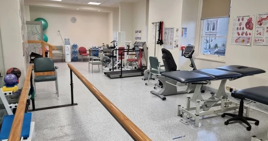
During recovery, the focus is on avoiding recurrences and ensuring joint stability.
The following procedures are performed during comprehensive rehabilitation:
- A course of physical therapy,
- Therapeutic massage,
- Special therapeutic exercises under the supervision of a specialist.


