Definition
Osteoarthritis of the ankle joint is a degenerative-dystrophic lesion of the joint’s cartilage plate and the underlying bone.
About the disease
The disease initially begins with damage to the cartilaginous base of the joint. Under the influence of unfavorable factors, the cartilage thins, loosens, and cracks, contributing to the exposure of the underlying bone. During movements in the joint, the exposed bone experiences an unphysiological load, so it tries to “protect” itself. It provokes compensatory osteosclerosis (thickening) in the underlying subchondral zone and the development of secondary subchondral cysts. In response, the ideal relationship of the articular surfaces is disturbed, further aggravating the pathologic process. As the disease progresses, the newly formed bone tissue at the edges forms overgrowths (osteophytes), which become the cause of intense pain syndrome.
Deforming osteoarthritis of the ankle can be triggered by various factors, including genetic, traumatic, growth, and metabolic conditions. The initial destruction of articular cartilage gradually leads to lesions of all synovial joint tissues.
Classification
According to the classification, two variants of the disease are distinguished:
- primary osteoarthritis, also called idiopathic, when the actual cause of the disease cannot be determined even with the most advanced examination;
- secondary osteoarthritis is due to exposure to a clearly delineated causative factor or factors listed above.
In clinical medicine, there are 6 degrees of ankle osteoarthritis:
- in the first degree – the superficial zone of cartilage is not damaged, but there is edema and matrix defibration, chondrocytes proliferate, and the type of collagen synthesized by them changes (in the normal cartilage plate is formed by collagen of the second type, but in osteoarthritis, it is replaced by less strong collagen of the third type);
- in the second degree, the integrity of the superficial zone of the cartilage plate is disturbed, and the location of chondrocytes in the deep zone is altered;
- In the third degree, the progression of the pathological process leads to the appearance of vertical cracks;
- in the fourth degree, the superficial zone of cartilage flakes off, eroded surfaces and cysts appear;
- the fifth degree is characterized by exposure of the underlying bone;
- at the sixth degree in the bone tissue, there are compensatory changes, which consist of its compaction, the formation of osteophytes, and microfractures.
Symptoms
The main manifestation of ankle osteoarthritis is pain. Distinctive characteristics of the pain syndrome in this disease are:
- the starting character of pain, when it is maximized at the onset of movement;
- mechanical nature, which leads to increased pain sensations during physical activity and during prolonged walking;
- nocturnal aching pain due to intraosseous venous blood stasis;
- blockage pain is a jam in the ankle, in which a person can neither bend nor straighten the leg, as the pain increases significantly (blockage occurs due to the jamming of fragments of dead cartilage between the articular surfaces);
Osteoarthritis of the ankle is a chronic process. Painful periods, which indicate an exacerbation of the disease, alternate with painless periods. With the progression of osteoarthritis, the inter-relapse period is shortened, and the pain may become permanent at a certain stage.
Causes of ankle osteoarthritis
On average, the cartilage plate gradually breaks down in people starting at 30, outpacing the rate of new cartilage formation. Therefore, the prevalence of the disease increases with age. There are also certain gender specificities. For example, before the onset of menopause, women’s joints are protected from destruction. With the onset of the menopausal transition, the protective effect of estrogen gradually decreases, so from the age of 50, the incidence of pathology in men and women is equalized.
The following causes of ankle osteoarthritis are distinguished, which lead to the fact that the process of cartilage re-synthesis does not manage to overlap with the catabolism of cartilage:
- Traumatic injuries (jumping on your feet from a height is hazardous);
- Previous inflammatory lesions of the joint;
- Ankle deformities, which may be associated with flat feet, varus or valgus;
- Hereditary collagenopathies, particularly those affecting type 2 collagen synthesis;
- Ankle dysplasia;
- Excessive body weight, which increases the load on the ankle and contributes to the “abrasion” of cartilage layers;
- Postmenopausal period (the average age of permanent cessation of menstruation in women is 50-52 years);
- Metabolic disorders;
- Sedentary lifestyle;
- Orthopedic interventions on the joint;
- Repeated hypothermia.
Diagnosis
If ankle joint osteoarthritis is suspected, the doctor recommends an additional examination program. It may consist of the following methods:
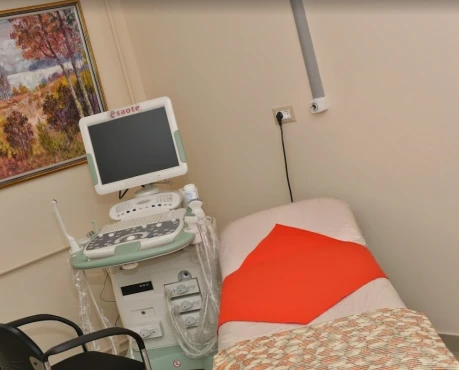
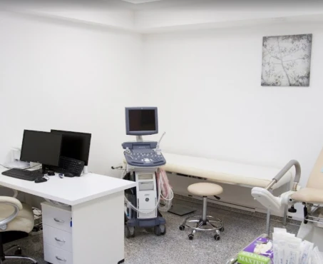
- ultrasound scanning – the study allows to assess the condition of soft tissue structures of the joint (cartilage, synovial bag, and surrounding tissues); it is the most informative method for early diagnosis of osteoarthritic changes;
- X-ray – this method primarily evaluates the structure of bone tissues, helps to detect subchondral osteosclerosis, the presence of cysts in the subcartilaginous zone, as well as to visualize osteophytes (with the help of radiography to detect the initial changes of osteoarthritis, mainly involving the cartilaginous plate, is extremely difficult).
In complex clinical cases, computed tomography or magnetic resonance imaging may be used to detail the condition of the ankle joint. Each method allows for obtaining layer-by-layer scans (scanning steps 2-3 mm) of the area under study and assessing the condition of the intra- and extra-articular structures of the ankle joint.
Treatment of ankle osteoarthritis
Conservative methods are used to treat osteoarthritis at its initial stages. Timely therapy can protect the joint from destruction and delay or avoid the need for surgical intervention. Endoprosthesis is indicated if the disease is detected at the stage of significant destruction of the cartilage plate and is accompanied by stiffness that disrupts human activity.
Conservative treatment
Conservative treatment of osteoarthritis begins with creating favorable conditions for joint functioning. Following measures are recommended:
- regular physical therapy, swimming, and aqua aerobics are also useful;
- normalization of body weight (in case of excess);
- use of crutches or orthopedic canes during the exacerbation of the process;
- wearing comfortable orthopedic shoes.
To improve the condition of the cartilage plate, chondroprotectors are used, which are injected mainly inside the joint. Hyaluronic acid and PRP therapy (plasma therapy) restore the condition of the cartilage plate. Symptomatic treatment with non-steroidal anti-inflammatory drugs is carried out to control pain.
Surgical treatment
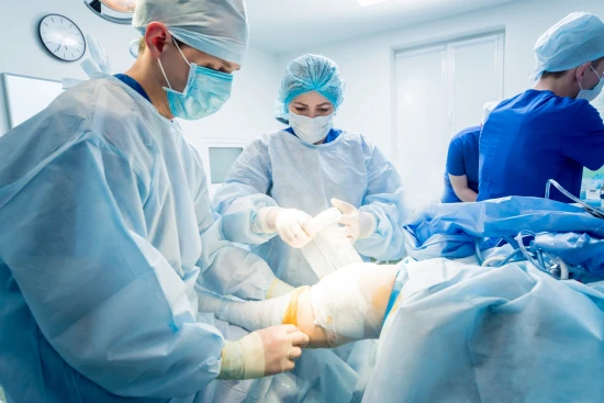
Prosthetics of the ankle joint is a rather complex task, so surgeons strictly follow the operation’s modern methodology, allowing them to achieve the best therapeutic results. Currently, only third-generation implants are used in this operation, which requires the removal of only a small bone fragment. These prostheses stimulate osteoclasts (bone-forming cells) and fuse well with the tibia, fibula, and talus, providing a solid structure. A unique feature of the third-generation prosthesis is that it allows movement not only of the main joint but also of the joint between the fibula and tibia, thus evenly distributing the load on the joint.
Ankle replacement surgery also involves correcting existing deformities and stitching damaged ligaments. It creates favorable conditions for maintaining the joint’s stability and ensuring its full function.
Prevention of ankle osteoarthritis
Prevention of ankle osteoarthritis is to follow the following recommendations:
- wearing comfortable and non-compressive footwear, using orthopedic insoles;
- doing as much exercise as you can;
- use of special ankle braces in professional sports;
- Eliminating foot jumps from heights;
- timely correction of concomitant deformities of the lower limb.
Rehabilitation
After the orthopedic intervention, the operated joint is temporarily immobilized. This period allows for optimal conditions for bone tissue regeneration and helps the implant integrate more fully. After the plaster cast is removed, recreational exercises under the supervision of a physical therapy doctor, massage, and physiotherapy are indicated.
