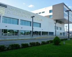
Patent foramen ovale (PFO) treatment in 1 Cardiac surgery and Vascular surgery clinic in Nicaragua

Top
Clinics
Diseases
Read about
Quick navigation

1 clinic specializing in Cardiac surgery and Vascular surgery providing treatment of Patent foramen ovale (PFO) 
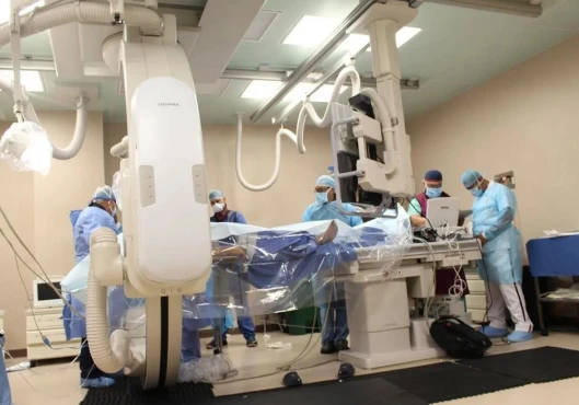
Patent foramen ovale (PFO) is a common congenital heart defect where a small hole between the upper chambers of the heart fails to close after birth. It can allow blood clots to pass from the right to the left side of the heart, potentially causing stroke or other complications.
Read more...
disease in Nicaragua.

Besides this clinic there is 1 Cardiac surgery, Vascular surgery clinic in Managua and 1 clinic in Nicaragua.

Such diseases are treated by Hospital Vivian Pellas: Atrial septal defect (ASD), Chronic thromboembolic disease, Chronic thromboembolic pulmonary hypertension, Intracardiac thrombus, Patent foramen ovale (PFO), and others.

Managua · 1
You can select a specific city
Managua · 1
Sorted by:
Relevance
Rating Relevance
Prices for popular procedures:
  1. Atrial septal defect (ASD) repair in children
    ≈ $6,963
  2. Percutaneous ASD closure
    ≈ $3,463
  3. NobleStitch™ PFO closure procedure
    by request
  4. Pulmonary angiography
    ≈ $888
  5. Dual chamber pacemaker insertion
    ≈ $5,787
  6. Carotid endarterectomy (CEA)
    ≈ $2,585
  7. Heart valve replacement
    ≈ $6,898
  8. Heart valve repair
    ≈ $6,271
  9. Aortic valve replacement (AVR)
    ≈ $12,174
photo
Managua, Nicaragua
Specializations: Cardiac surgery, Vascular surgery, Thoracic surgery, Neurosurgery, Spine surgery, Orthopedic surgery, Oncology
The Vivian Pellas Hospital opens its doors with the vision of being the organization that raises the standard of private health in Nicaragua to the read more

1 nearby similar clinic in Nicaragua

We found only 1 clinic in Nicaragua that matches your criteria, presented in the list above.
Perhaps you should consider 1 more clinic we have found nearby basing on your Location, Specialization, Disease filters applied.
Prices for popular procedures:
  1. Arteriovenous (AV) fistula surgery
    ≈ $757
  2. Endovascular thrombectomy
    ≈ $2,719
  3. Coronary catheterization
    ≈ $756
  4. Percutaneous coronary intervention (PCI) with stent insertion
    ≈ $5,736
  5. Percutaneous coronary intervention (PCI) with angioplasty
    ≈ $2,644
  6. Fractional flow reserve (FFR)
    ≈ $573
  7. Rotational atherectomy
    ≈ $896
  8. Reconstructive surgery for peripheral artery disease (PAD)
    ≈ $1,717
  9. Arteriovenous (AV) graft surgery
    ≈ $1,110
photo
Managua, Nicaragua
Specializations: Cardiac surgery, Vascular surgery, Thoracic surgery, Neurosurgery, Orthopedic surgery, Oncology
We are a hospital with the capacity to provide third and fourth level medical care services to members of the Nicaraguan Army. We have highly read more

Countries with the highest number of clinics treating the diseases:

Patent foramen ovale (PFO):

Related procedures:

Patent foramen ovale (PFO) as one of the conditions of minor heart anomalies

Hemodynamic disturbances in minor heart anomalies

Our body is a unique system in which scientists are not always able to draw a clear line between the norm and pathology. One such situation is when a patient has one or more conditions from the so-called group of minor heart anomalies (MHA). In this article, we will analyze in detail what these defects are and what features of hemodynamics (blood flow) they have.

Why was the term "minor heart anomalies" used?

Improving the diagnosis of heart diseases, the accumulation and analysis of the results of scientific research and databases have led to the interest of doctors and scientists in borderline changes in the structure of the heart. Some authors refer to the latter as MHA. However, this term is not international, and the states that can be attributed to it remain a matter of debate.

MHA belongs to connective tissue dysplasia (CTD) — a clinically heterogeneous group of diseases that are caused by primary (genetic) or secondary (non-genetic) disorders in the production and/or breakdown of connective tissue proteins and components of the extracellular substance.

Various pathologies can be attributed to MHA, however, in this article, in the context of MHA, we will analyze hemodynamics in the following conditions:

  1. mitral valve prolapse (MVP);
  2. aneurysm of the interatrial septum (AIS);
  3. patent foramen ovale (PFO);
  4. an elongated Eustachian valve (EV);
  5. Chiari networks.

However, before the main part of the article, it is necessary to recall important aspects of the anatomy of the heart and hemodynamics in normal conditions. So, the heart consists of four cavities: two ventricles and two atria. The left side of the heart muscle pumps blood through the systemic circulation, and the right side through the small one. Both halves are separated by a partition. An important role in proper hemodynamics is played by the valves of the heart, which prevent the reverse flow of blood.

Mitral valve prolapse (MVP)

MVP is detected by ECHO-CG in 2-3% of patients from the general population. The main complications of this pathology are associated with the progression of mitral insufficiency. When it occurs, the reverse flow of blood (regurgitation) from the left atrium into the left ventricle. Also, this pathology increases the risk of endocarditis (inflammation of the inner layer of the heart - endocardium), sudden cardiac death and stroke.

Elongated Eustachian valve (EV)

The remnants of the Eustachian valve (EV) are parts of the embryological valves of the venous sinus (it is a rudimentary, anatomically variable structure). The average length of the EC is 3.6 mm with a range of 1.5 to 23 mm. It may persist as a mobile oblong structure extending into the right atrium. Thus, it can be mistaken for an abnormal formation in the heart. The Eustachian valve should be differentiated from atrial tumors, thrombi, or vegetations.

A large EV may be associated with impaired hemodynamics due to the direction of blood flow through the inferior vena cava into the left atrium through an atrial septal defect. It may also predispose to paradoxical embolism in patients with patent foramen ovale by the same mechanism. A large EV can cause an obstruction (blockage) in the inferior vena cava. Myxoma, papillary fibroelastoma and cyst can also occur from it. In some cases, EV causes endocarditis, pulmonary thromboembolism and provokes the occurrence of cardiac arrhythmia.

The manifestations and clinical significance of an elongated EV are determined by the association with other anomalies, difficulties in differential ECHO-CG diagnostics, and possible complications.

Chiari Network

The Chiari network is a congenital anatomical variation at the junction of the right atrium with the superior and inferior vena cava. The network was first described in the medical literature in 1875.

The prevalence of the Chiari network varies from 1.3 to 4% with post-mortem findings and from 0.3 to 9.5% with transthoracic echocardiography. This pathology may also be associated with an increased prevalence of other congenital anomalies, including patent foramen ovale (PFO) and atrial septal aneurysm. However, there was no convincing evidence that this defect can interfere with blood flow.

The medical literature describes cases of difficulty in passing catheters into various structures of the heart if the patient has a Chiari network.

Patent foramen ovale (PFO)

In 75% of the population, the foramen ovale spontaneously closes normally at birth with an increase in left atrial pressure and a decrease in pulmonary resistance. However, it persists as an open pulmonary collar in 15–35% of the adult population. A patent foramen ovale is an embryonic atrial septal defect that allows oxygenated blood to pass from the right atrium to the left atrium. The PFO is of clinical importance because it can be a source of thrombus formation or serve as a conduit for paradoxical embolism.

Atrial septal aneurysm

This condition is a local atrial septal defect, consisting of its excess and mobile tissue. This defect is located in the region of the oval fossa and protrudes into the right or left atrium, and sometimes fluctuates between both atria. Although its prevalence in the general population has not been reported, the defect is thought to occur in 2–3% of the population. Except for its potential role in cardioembolic stroke and its structural association with valvular regurgitation, patent foramen ovale, and atrial septal defect, it has been considered an incidental and clinically asymptomatic finding in adult patients.

Thus, clinically significant hemodynamic disturbances in minor cardiac anomalies are quite rare. Most often, these conditions are associated with regurgitation, an increased risk of thrombosis and endocarditis. It is not uncommon for different forms of MHA to be found in the same patient.

References:

  1. Basso C, Iliceto S, Thiene G, Perazzolo Marra M. Mitral Valve Prolapse, Ventricular Arrhythmias, and Sudden Death. Circulation. 2019;140(11):952-964.
  2. Althunayyan A, Petersen SE, Lloyd G, Bhattacharyya S. Mitral valve prolapse. Expert Rev Cardiovasc Ther. 2019;17(1):43-51.
  3. Gulel O, Yazici M, Sahin M. Unusual elongation of the Eustachian valve. Int Heart J. 2007;48(1):113-116.
  4. Teshome MK, Najib K, Nwagbara CC, Akinseye OA, Ibebuogu UN. Patent Foramen Ovale: A Comprehensive Review. Curr Probl Cardiol. 2020;45(2):100392.
  5. Yetkin E, Ileri M, Korkmaz A, Ozturk S. Association between atrial septal aneurysm and arrhythmias. Scand Cardiovasc J. 2020;54(3):169-173.
  6. Supranational (international) recommendations for structural anomalies of the heart // Medical Bulletin of the North Caucasus. 2018. No. 1.2.

Quick navigation