Introduction
The ankle joint is the most congruent joint of the lower limb, and the bones’ cartilage forming it is characterized by the smallest thickness. A defect in the cartilage of the talus bone is a consequence of traumatic damage to the ankle joint. This pathology is accompanied by pain syndrome and impaired movement in the ankle joint. Cartilage damage can occur at any age. Most cases of this problem result from an ankle injury with a torsional component and is essentially a fracture of the articular surface of the talus bone.
The incidence of osteochondral defects of the talus in patients with fresh ankle injuries is 7-9%. These defects cause long-lasting swelling, limited range of motion, and pain on loading. The defect may be visible on radiographs, but more often, they are uninformative. Traumatic osteochondral lesions are characterized by low regenerative capacity and, therefore, require special surgical treatment.
There are also cartilage defects in the ankle joint, the causes of which are not directly related to trauma. Here, blood circulation in the bone may be impaired due to necrosis and genetic factors. Most of them are asymptomatic for a long time. However, they may later manifest themselves with pain and swelling, for example, after an episode of trauma.
Osteochondral defects are usually localized in the anterior part of the talus block. They often result in the formation of loose bodies in the ankle joint. Repeated overloading of the damaged articular cartilage can lead to further localized cartilage cell death.
Reasons
A common cause of cartilage damage is a fracture of the articular surface of the talus. It is usually combined with damage to the ankle ligaments. The subsidence of the articular surface can vary considerably in shape and area depending on the severity of the traumatic injury. In cases of severe damage to the cartilage and adjacent bone, the damaged fragment may detach, forming a loose articular body in the joint (“articular mouse”).
Due to the complete detachment of the bone fragment and cartilage, its blood supply is disrupted. As a result, necrosis of the tissue of the displaced fragment is noted. This variant explains those cases in which the patient has no history of severe ankle trauma with potential fracture development.
Classification of osteochondral defects of the talus
Stage I: minor compression fracture.
Stage II: incomplete detachment of the fragment.
Stage III: complete detachment of the fragment with its displacement.
Stage IV: displaced fragment.
Symptoms
Initially, patients feel a sharp pain that increases with support on the affected leg. X-rays of the ankle joint are taken to confirm the diagnosis. In the late period, patients still have pain when walking and swelling in the ankle joint. In the case of a free fragment in the joint cavity, there may be a crunch when moving and a feeling of “joint blockage.” Patients with cartilage damage usually experience pain deep in the ankle joint during or after physical activity. The cause of the pain syndrome is the increased pressure inside the ankle during walking, which irritates the bone pain receptors at the bottom of the cartilage defect.
Diagnosis
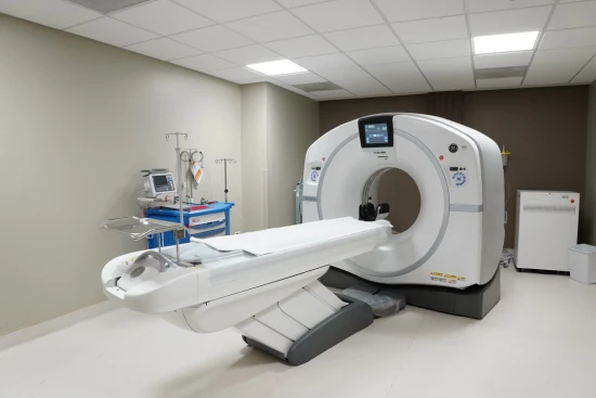
An ankle cartilage defect can be diagnosed based on history and examination. Ankle radiographs usually confirm a fracture of the articular surface of the talus. In some cases, magnetic resonance imaging and computed tomography are ordered to confirm the diagnosis.
Treatment
Conservative treatment
Asymptomatic or minimally manifested injuries are treated conservatively: rest, ice, temporary load limitation, and, if the joint feels unstable, the use of special orthoses.
In the acute period, immobilizing the ankle joint with a plaster cast or special fixator is recommended for up to 6 weeks. During this period, it is necessary to use crutches when walking. In the first days after the injury, an elevated position of the lower extremity, cold packs, and non-steroidal anti-inflammatory drugs are indicated to reduce the severity of pain syndrome and local edema.
If symptoms persist for several months after the injury and a free bone and cartilage fragment is detected in the joint cavity, surgical intervention is necessary – ankle arthroscopy. It consists of removing the free body and tunnelization or microfracturing of the damaged area of bone. This procedure improves blood flow in the area of the injury. Over time, scar tissue forms in the area of detached cartilage, which entirely or partially closes the defect. As a result, the articular surface of the talus is restored, and the movement volume is restored.
Surgical treatment
Using arthroscopy, this operation can be performed without incisions. The arthroscope is an optical system consisting of a miniature video camera, an illuminator, and a rigid light guide. It is inserted into the joint cavity through a puncture and allows inspection of the interior of the joint. Free-body removal and tunnelization or microfracturing of the bone can be performed with miniature instruments.
During an ankle arthroscopy, debridement and bone marrow stimulation are usually performed. The technique consists of removing all detached fragments of cartilage and the underlying necrotized bone. If there are cysts in the bone under the cartilage, they are opened and treated uniquely. The bone is then microperforated with a special instrument. It leads to the release of growth factors that fill the cartilage defect in the talus. The formation of new blood vessels is stimulated, and stem cells from the bone marrow of the talus are released into the osteochondral defect, which is subsequently filled with cartilage.
During surgery, intra-articular pressure on the cartilage is reduced, which blocks the stimulation of nerve endings and reduces the severity of pain in the patient. Good and excellent results are noted in 86% of patients.
Rehabilitation
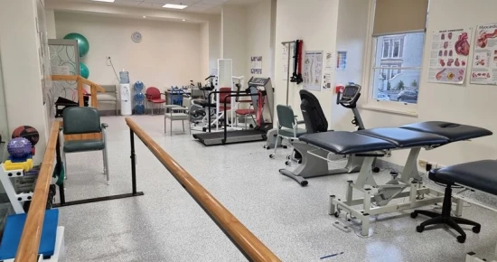
After surgery, crutches should be used for 3-6 weeks to relieve pressure on the operated limb. From the first days, the restoration of the range of motion begins. For this purpose, special exercises are prescribed to increase the joint’s movement amplitude. To reduce edema and postoperative pain syndrome, physiotherapy (magnetotherapy, electroanalgesia, laser therapy, etc.) or shockwave therapy may be recommended.


