Kohler's disease (accessory navicular syndrome, osteochondropathy of the navicular bone of the foot) is a degenerative bone disease of the foot. It is characterized by impaired blood supply to the navicular bone, which can lead to degeneration and destruction of the joint. Osteochondropathy of the navicular bone of the foot usually develops during the adolescent period of rapid growth, and its symptoms may include pain in the midfoot and forefoot, swelling, limitation of motion, and limping. With long-term progression of the condition, foot deformities such as flat feet or clubfoot may occur.
Causes of Kohler disease
The causes of Kohler disease can be outlined in several points:
- increased stress on the foot, including intense sports training;
- rapid growth and development during adolescence;
- genetic predisposition;
- mechanical factors such as improper foot placement.
Risk factors
Risk factors for accessory navicular syndrome are defined by these characteristics:
- intense sports training, especially those involving jumping and other intense physical stresses on the foot;
- rapid growth and development during adolescence;
- genetic predisposition;
- improper foot placement;
- increased stress on the foot due to long-term standing or walking on hard surfaces such as concrete floors or asphalt.
Pathogenesis
The pathogenesis of osteochondropathy of the navicular bone of the foot can be characterized as:
- impaired blood supply, which can be caused by vasoconstriction or damage to the microcirculation;
- damage or destruction of cartilage tissue, resulting in impaired normal development and growth;
- inflammatory processes that may occur in response to trauma or microtrauma;
- changes in metabolism, such as impaired metabolism of calcium, phosphorus, and other trace minerals that affect bone health;
- genetic factors, including inherited mutations that can affect bone structure and function.
Symptoms of accessory navicular syndrome
Symptoms and signs of Kohler disease in an adult can be identified as follows:
- pain in the plantar part of the foot, especially on exertion;
- swelling and inflammation;
- limitation of the mobility of the foot;
- hypersensitivity or numbness;
- deterioration of foot function, difficulty walking;
- increased pain when bending the foot or moving on uneven ground;
- there may be a visible change in the shape of the foot or a bulge in the head of the navicular bone;
- pain may increase during physical activity and decrease at rest.
In some cases, the foot movement may be restricted due to aseptic necrosis and discomfort.
Complications
The following indicators identify complications of Kohler disease:
- inflammation can spread to the other foot;
- swelling;
- pain;
- traffic restriction;
- deformation;
- increased risk of fractures;
- foot dysfunction;
- limiting physical activity;
- gait disturbance;
- decreased quality of life;
- Long-term restrictions on daily activities.
Diagnosis of Kohler disease
Diagnosing Kohler disease of the foot involves performing medical manipulations:
- gathering medical history: information about symptoms, duration of pain, previous injuries or surgeries, and presence of comorbidities;
- clinical examination of the foot and lower leg: assessing range of motion, checking for pain on palpation, and evaluating foot structure and function;
- X-ray: anteroposterior and lateral views to visualize bone structure and identify possible changes inherent in osteochondropathy;
- magnetic resonance imaging (MRI) of the foot, to visualize soft tissues, ligaments, joints, and bones in more detail and to detect early stages of osteochondropathy;
- bone scanner densitometry to assess bone density and identify possible osteoporosis that may be associated with osteochondropathy;
- laboratory tests, such as blood tests for inflammatory markers or biochemical indicators, which can help rule out other possible causes of foot pain;
- consultation with specialists: orthopedist or rheumatologist, for additional opinion and determination of optimal treatment;
After carrying out all the necessary diagnostic procedures, the doctor can accurately diagnose the pathology and prescribe appropriate treatment.
Treatment of accessory navicular syndrome
Treatment of accessory navicular syndrome includes the following approaches:
- Corticosteroids: the use of corticosteroid medications such as prednisolone, methylprednisolone, or dexamethasone can reduce inflammation and symptoms of Kohler disease;
- Immunosuppressants: azathioprine, methotrexate, or cyclosporine can be used to suppress the immune system and reduce inflammation;
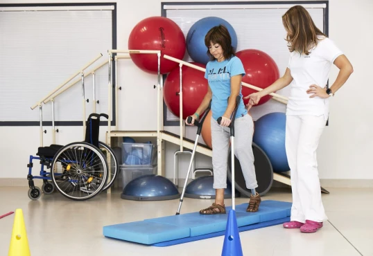
- Physical therapy: physical therapy can help improve joint mobility and relieve pain; physical therapy techniques include joint exercises, medical massage, ultrasound therapy, and other treatments;
- Pain medications: Non-steroidal anti-inflammatory drugs (NSAIDs) such as ibuprofen or diclofenac can be used to relieve pain and reduce inflammation;
- Orthopedic shoes and orthoses: the use of special orthopedic shoes and orthoses to support and stabilize affected joints, improve walking, and reduce strain;
- Good nutrition: good nutrition helps to strengthen the immune system and improve the general condition of the body;
- Regular follow-up with a doctor can monitor the condition of the disease, regulate treatment, and detect possible complications in time.
Surgery for Kohler disease
Surgery may be required if conservative treatment does not result in sufficient improvement. However, the doctor determines the need for surgery and its type based on the disease's severity and the patient's individual characteristics.
Surgery for Kohler disease may include the following options:
- Arthrodesis: this procedure stabilizes the joint by fixing it using an implant or other methods; it may be recommended when the joint is severely damaged and no other treatment options are available;
- Metatarsal head resection: the upper part of the metatarsal bone (head) is removed to reduce pressure on the joint and improve its function;
- Arthroplasty: in some cases, a joint may be replaced with an implant to restore functionality and reduce pain;
- other procedures: depending on the specifics of the disease and the patient's medical history, other surgical interventions such as correction of foot deformities, synovectomy (removal of the inflamed synovial membrane of the joint), Kidner procedure etc. may be recommended.
It is important to note that an orthopedic surgeon should decide whether to perform surgery and what type of surgery to perform based on a comprehensive evaluation of the patient's condition, examination results, and medical history. After surgery, rehabilitation may be required, and the doctor's recommendations may need to be followed for optimal recovery results.
Prognosis
The prognosis and consequences of the disease are as follows:
- continued pain;
- possible progression of the deformity;
- outcome: deterioration of foot function;
- limiting physical activity;
- possible complications such as inflammation and swelling;
- need for long-term treatment and rehabilitation;
- decreased quality of life;
- possible limitations of movement in everyday life;
- need for constant supervision and monitoring by a physician.
Prevention of Kohler disease
Prevention of Kohler disease involves:
- wearing comfortable and appropriate footwear;
- controlling the load on the foot during physical activity;
- regular exercises to strengthen the muscles of the foot;
- avoiding prolonged standing and walking on hard surfaces;
- maintaining a healthy lifestyle, including proper nutrition;
- timely referral to a doctor in case of pain and discomfort in the foot;
- compliance with the doctor's recommendations for the treatment and prevention of osteochondropathy of the navicular bone of the foot;
- avoiding traumatic impacts on the foot, such as bruises and fractures;
- regular preventive check-ups with a doctor for early detection of changes in the foot;
- use of orthotic insoles or pads, if necessary, to reduce stress on the foot.


