Ventricular septal defect (VSD) treatment in 1 Cardiac surgery clinic in Slovakia

Top
Clinics
Diseases
Read about
Quick navigation

1 clinic specializing in Cardiac surgery providing treatment of Ventricular septal defect (VSD) Ventricular septal defect (VSD) is a congenital heart defect where there is a hole in the wall (septum) separating the heart's lower chambers (ventricles). It can lead to abnormal blood flow and heart strain, requiring medical monitoring and, in some cases, surgical repair in early childhood.
Read more...
disease in Slovakia.

Besides this clinic there is 1 Cardiac surgery clinic in Bratislava and 3 clinics in Slovakia.

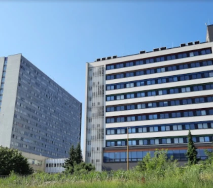
Such diseases are treated by National Institute for Cardiovascular Diseases, Bratislava: Acute ST-elevation myocardial infarction (STEMI), Atrioventricular canal defect, Pulmonary stenosis, Transposition of the great arteries (TGA), Ventricular septal defect (VSD), and others.

You can select a specific city
Sorted by:
Relevance
Rating Relevance
Prices for popular procedures:
  1. Pulmonary artery banding (PAB)
    ≈ $7,199
  2. Ventricular septal defect (VSD) repair in children
    ≈ $5,911
  3. Ventricular septal defect (VSD) repair
    ≈ $11,671
  4. Percutaneous closure of ventricular septal defect (VSD)
    ≈ $6,431
  5. Nikaidoh procedure
    ≈ $9,182
  6. Coronary artery bypass graft (CABG)
    ≈ $10,479
  7. Heart valve replacement
    ≈ $11,534
  8. Heart valve repair
    ≈ $11,963
  9. Aortic valve replacement (AVR)
    ≈ $11,994
photo
Bratislava, Slovakia
Specializations: Cardiac surgery, Vascular surgery
The National Institute of Heart and Vascular Diseases is one of the most modern and best-equipped Slovak hospitals, the top institution of cardiovascular medicine in read more

Nearby clinics in Slovakia

We found only 1 clinic in Slovakia that matches your criteria, presented in the list above.
Perhaps you should consider the following clinics we have found nearby basing on your Location, Disease filters applied.
Prices for popular procedures:
  1. Percutaneous coronary intervention (PCI) with stent insertion
    ≈ $4,967
  2. Fractional flow reserve (FFR)
    ≈ $615
  3. Percutaneous coronary intervention (PCI) with angioplasty
    ≈ $3,355
  4. Rotational atherectomy
    ≈ $1,047
  5. Pulmonary artery thromboendarterectomy
    ≈ $6,374
  6. Arteriovenous (AV) fistula surgery
    ≈ $679
  7. Endovascular thrombectomy
    ≈ $2,550
  8. Varicose veins treatment
    ≈ $1,067
  9. Unilateral varicose veins surgery
    ≈ $666
photo
Ružomberok, Slovakia
Specializations: Cardiac surgery, Vascular surgery, Neurosurgery, Spine surgery, Orthopedic surgery, Oncology
The Central Military Hospital of the SNP Ružomberok provides its services to all citizens of the Slovak Republic who need it, regardless of where they read more
Prices for popular procedures:
  1. Angioplasty of the pulmonary artery
    ≈ $2,045
  2. Pulmonary artery thromboendarterectomy
    ≈ $6,374
  3. Abdominal endovascular aneurysm repair (EVAR)
    ≈ $10,109
  4. Thoracic endovascular aneurysm repair (TEVAR)
    ≈ $9,562
  5. Peripheral artery angioplasty and stent placement
    ≈ $2,986
  6. Renal artery angioplasty and stenting
    ≈ $2,581
  7. Arteriovenous (AV) fistula surgery
    ≈ $679
  8. Endovascular thrombectomy
    ≈ $2,550
  9. Uterine artery embolization (UAE)
    ≈ $2,506
photo
Martin, Slovakia
Specializations: Cardiac surgery, Vascular surgery, Thoracic surgery, Neurosurgery, Spine surgery, Orthopedic surgery, Oncology
The Martin University Hospital is a terminal health care facility of supra-regional importance with several exceptional workplaces with nationwide scope. An average of 50 percent read more
Prices for popular procedures:
  1. Dual chamber pacemaker insertion
    ≈ $5,220
  2. Permanent pacemaker implantation
    ≈ $4,137
  3. Removal of permanent pacemaker
    ≈ $2,790
  4. Single chamber pacemaker insertion
    ≈ $4,677
  5. Cardiac resynchronization therapy with pacemaker (CRT-P)
    ≈ $7,359
  6. Cardiac resynchronization therapy with defibrillator (CRT-D)
    ≈ $9,495
  7. Pacemaker replacement
    ≈ $1,966
  8. Temporary pacemaker implantation
    ≈ $500
  9. Leadless Pacemaker implantation Micra and Nanostim
    ≈ $5,996
photo
Bratislava, Slovakia
Specializations: Cardiac surgery, Vascular surgery, Thoracic surgery, Neurosurgery, Spine surgery, Orthopedic surgery, Oncology
Languages: English
Nemocnica Ružinov is the largest hospital in Slovakia. University Hospital Bratislava (UNB) is the largest medical facility in Slovakia. The basic mission of UNB is read more
Prices for popular procedures:
  1. Pulmonary artery thromboendarterectomy
    ≈ $6,374
  2. Arteriovenous (AV) fistula surgery
    ≈ $679
  3. Endovascular thrombectomy
    ≈ $2,550
  4. Radiofrequency ablation (RFA) for varicose veins (Unilateral)
    ≈ $991
  5. Endovenous laser ablation (EVLA) for varicose vein (Unilateral)
    ≈ $1,058
  6. Varicose vein stripping (Unilateral)
    ≈ $1,043
  7. Phlebectomy (including mini-phleboctomy)
    ≈ $646
  8. Catheter-directed sclerotherapy (CDS) (Unilateral)
    ≈ $265
  9. Foam sclerotherapy (Unilateral)
    ≈ $192
photo
Bratislava, Slovakia
Specializations: Cardiac surgery, Vascular surgery, Thoracic surgery, Neurosurgery, Spine surgery, Orthopedic surgery, Oncology
Ladislav Dérer Hospital (Kramáre Hospitalis) is a hospital in the New Town district of Bratislava, local part of Kramáre. Its old building was built in read more
Prices for popular procedures:
  1. Pulmonary artery thromboendarterectomy
    ≈ $6,374
  2. Arteriovenous (AV) fistula surgery
    ≈ $679
  3. Endovascular thrombectomy
    ≈ $2,550
  4. Carotid endarterectomy (CEA)
    ≈ $2,870
  5. Carotid angioplasty and stenting
    ≈ $4,409
  6. Varicose veins treatment
    ≈ $1,067
  7. Unilateral varicose veins surgery
    ≈ $666
  8. Radiofrequency ablation (RFA) for varicose veins (Unilateral)
    ≈ $991
  9. Endovenous laser ablation (EVLA) for varicose vein (Unilateral)
    ≈ $1,058
photo
Bratislava, Slovakia
Specializations: Cardiac surgery, Vascular surgery, Thoracic surgery, Neurosurgery, Spine surgery, Orthopedic surgery, Oncology
University Hospital Bratislava - St. Cyril and Methodius Hospital is a hospital in the Petržalka district of Bratislava , in its local part of read more
Prices for popular procedures:
  1. Angioplasty of the pulmonary artery
    ≈ $2,045
  2. Pulmonary artery thromboendarterectomy
    ≈ $6,374
  3. Peripheral artery angioplasty and stent placement
    ≈ $2,986
  4. Renal artery angioplasty and stenting
    ≈ $2,581
  5. Endovascular thrombectomy
    ≈ $2,550
  6. Uterine artery embolization (UAE)
    ≈ $2,506
  7. Varicose veins treatment
    ≈ $1,067
  8. Unilateral varicose veins surgery
    ≈ $666
  9. Radiofrequency ablation (RFA) for varicose veins (Unilateral)
    ≈ $991
photo
Bratislava, Slovakia
Specializations: Cardiac surgery, Vascular surgery, Orthopedic surgery, Oncology
Staré Mesto Hospital is part of the University Hospital Bratislava (UNB) is the largest medical facility in Slovakia. The basic mission of UNB is to read more

Countries with the highest number of clinics treating the diseases:

Ventricular septal defect (VSD):

Related procedures:

Ventricular septal defect (VSD) – disease overview

Origin and mechanisms of development of a ventricular septal defect

Ventricular septal defect (VSD) is one of the most common congenital heart defects. Its frequency is approximately 300 cases per 100,000 births. Also, VSD accounts for about a third of all congenital heart defects. Females and males get sick with the same frequency.

This disease was first described in 1879, and more than 100 years later, in 1988, the first percutaneous intervention was performed to close a defect in the interventricular septum.

Below we will analyze in detail the origin and mechanisms of development of the interventricular septal defect, and also touch on the treatment and diagnosis of this pathology in both children and adults.

Recall that the heart's main function is to pump blood throughout the body. This organ has two phases of its activity: contraction (systole) and relaxation (diastole).

The human heart consists of four cavities (two ventricles and two atria) and has four valves that prevent blood from flowing back. The latter is normally sent from the atria to the ventricles, and from them to the vessels (to the aorta from the left cavity, and to the pulmonary arteries from the right). The right ventricle (RV) is separated from the left ventricle (LV) by a septum, which consists of a muscular and membranous portion.

The pathogenesis of ventricular septal defect and its complications is inextricably linked with hemodynamics (direction of blood flow). The LV has a massive muscle mass and creates pressure several times higher than that in other chambers of the heart, therefore, when an opening in the interventricular septum is formed, blood is directed from left to right.

Causes of the disease and risk factors for ventricular septal defect

The exact cause of the ventricular septal defect is currently unknown, but congenital heart defects appear to be inherited and sometimes co-occur with other genetic diseases and chromosomal abnormalities such as Down syndrome.

Certain conditions that the mother already has or that occur during pregnancy can also increase the risk of the disease and cause VS defects. These include:

  1. Infection with rubella during the first few months of pregnancy.
  2. Use of drugs, tobacco, alcohol or exposure to other toxic substances.
  3. Diabetes mellitus (including those arising during pregnancy).
  4. Systemic lupus erythematosus.
  5. Overweight (body mass index over 25).
  6. Radiation exposure (diagnostic procedures such as radiography are not included in this item).
  7. Phenylketonuria.

VSD classification

This pathology is classified depending on the location of the pathological opening. So, the following types of ventricular septal defect are distinguished:

  1. Perimembranous (occurs in about 80% of cases) - the hole is located in the membranous part of the cardiac septum.
  2. Muscular (15–20%) - located in the muscular part of the septum and does not border on the fibrous rings.
  3. Subpulmonary outflow tract defect (about 5%) - borders on both fibrous rings of arterial valves.
  4. Defect of the bringing tract - contacts from above with the ring of the tricuspid (tricuspid) valve and is located behind the membranous septum.

Also, there is a large and small congenital malformation of the IVS. The size of the first is larger than the diameter of the aorta, the size of the second is smaller than the aortic lumen.

Is VSD a pathology in adults or children?

VSD is most commonly diagnosed and treated in children. However, the disease also occurs in adults and can be in the form of:

  1. A defect closed in childhood or a small unoperated defect resulting from a minor left-right shunt. Most often, a patient with such a defect does not have VSD symptoms, and due to the absence of left ventricular overload, surgery is usually not required.
  2. A defect that led to left ventricular overload and pulmonary hypertension. A consequence of the latter is a disease of the pulmonary vessels (Eisenmenger's syndrome).

Disease manifestations

A small VSD usually does not cause symptoms. For a more serious defect, signs of the disease include:

  • shortness of breath on exertion;
  • decreased exercise tolerance;
  • heartbeat.

In a newborn, as in an adult, symptoms may be absent with a small defect. There may be a murmur over the heart in more severe pathology. A significant defect is manifested by heart failure in a child aged 4–6 years. Symptoms in young children may include:

  • difficulties with feeding, poor weight gain, mental and physical retardation;
  • shortness of breath, rapid breathing and palpitations;
  • enlarged liver (hepatomegaly);
  • recurrent respiratory tract infections.

Why is this pathology dangerous?

Like other defects of the cardiovascular system, VSD without treatment (much less often if present) can pose a threat to the patient's life. The disease can have the following complications:

  • pulmonary hypertension;
  • Eisenmenger's syndrome;
  • infection of the inner layer of the heart (endocarditis);
  • left ventricular heart failure;
  • subvalvular aortic stenosis and other acquired heart defects.

Diagnosis of the disease

The doctor may suspect the presence of the disease immediately after childbirth or even during pregnancy (depending on the severity of the pathology). If there are complaints, the doctor collects all the necessary information and conducts an examination.

On auscultation (listening to murmurs), the doctor may find:

  1. Diastolic murmur, best of all audible in the III-IV left intercostal space at the sternum (characteristic symptom).
  2. Systolic tremor in the IV left intercostal space at the sternum, bifurcation of the II tone above the pulmonary artery.

The following instrumental diagnostic methods can be used:

  1. ECG. On the record, the doctor can detect signs of hypertrophy (enlargement) of the left atrium and ventricle, sometimes an incomplete block of the right bundle branch (block of one of the branches of the heart network, through which an excitation pulse passes, leading to a heart contraction).
  2. Plain X-ray of the chest organs. An increase in the vascular pattern of the lungs and an expansion of the heart contour can be detected.
  3. Echocardiography. It is the main method of visualization of pathology, which makes it possible to assess the location and size of the defect, its complications and accompanying anomalies. Also, the study reveals the relationship between pulmonary and systemic blood flow (usually this ratio is denoted by Qp: Qs).
  4. Magnetic resonance imaging (MRI) and computed tomography (CT) are used when echocardiography has not revealed pathology.
  5. Cardiac catheterization. Performed for the diagnosis of pulmonary hypertension and before treatment with the invasive VSD method.

Treatment: therapy in adult patients

Surgical treatment is usually not required for a small defect that is not accompanied by left ventricular overload, that is, in the case of an asymptomatic patient.

Surgery, regardless of symptoms, is performed in case of heart overload. The possibility of surgical treatment is also being considered in the following cases:

  • recurring episodes of endocarditis and a significant amount of blood leakage from the left ventricle;
  • progressive insufficiency of the aortic valve;
  • an increase in pulmonary pressure, at which there is a significant blood flow from left to right (Qp: Qs more than 1.5).

Treatment: therapy of VS defect in children

Isolated VSD in a newborn usually does not require treatment due to the possibility of spontaneous closure (especially if the defect is small and isolated). Therefore, only observation is often carried out.

In young children, the following surgical methods of treatment are used:

  • surgical closure of the defect;
  • percutaneous placement of an implant covering the defect;
  • pulmonary artery ligation - performed only as a palliative treatment when surgery is not possible.

Risks of surgical treatment

Treatment of this condition is usually successful and carries a very low risk of death. Most children will not experience major complications after surgery. There is a small chance that the patient may need re-intervention.
The surgery has a small risk of bleeding, blood clots inside the heart, and infection.

Disease prognosis

The disease in most cases is treatable or, as noted earlier, goes away with age. An unfavorable prognosis is observed only in persistent pulmonary hypertension in patients after defect closure.

Summary

Ventricular septal defect (VSD) is one of the most common congenital heart defects. A small VSD usually does not cause symptoms. For more serious malformation in adults, symptoms include exertional dyspnea, decreased exercise tolerance, and increased heart rate.

Surgical treatment is usually not required for a small defect that is not accompanied by left ventricular overload, that is, in the case of an asymptomatic patient. In other cases, the anomaly needs to be closed. In this case, both open and percutaneous low-traumatic methods can be used, which have minimal risks.

References:

  1. Harrison`s Principles of Internal Medicine 19/E (Vol.1). Dennis Kasper, Anthony Fauci, Stephen Hauseret all. McGraw-HillEducation 2015 ISBN: 0071802134 ISBN-13(EAN): 9780071802130.
  2. Interna szczeklika - duży podręcznik. Medycyna praktyczna. 2021. ISBN 9788374306522.
  3. Internal diseases in 2 volumes: textbook / Ed. ON THE. Mukhina, V.S. Moiseeva, A.I. Martynova - 2010 .-- 1264 p.
  4. Shlyakhto, EV Cardiology: national leadership / ed. E. V. Shlyakhto - 2nd ed. , revised and add. - Moscow: GEOTAR-Media, 2019 .-- 800 p. (Series: National Guide) - ISBN 978-5-9704-4810-6.

Quick navigation