Tetralogy of Fallot (TOF) treatment in 1 Cardiac surgery clinic in Guangzhou

Top
Clinics
Diseases
Read about
Quick navigation

1 clinic specializing in Cardiac surgery providing treatment of Tetralogy of Fallot (TOF) Tetralogy of Fallot (TOF) is a congenital heart defect characterized by four abnormalities: ventricular septal defect, pulmonary stenosis, overriding aorta, and right ventricular hypertrophy. It causes cyanosis and requires surgical correction in infancy.
Read more...
disease in Guangzhou.

Besides this clinic there are 7 Cardiac surgery clinics in China.

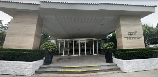
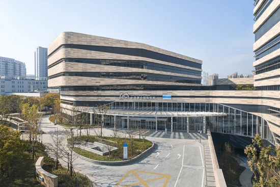
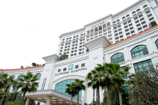
Such diseases are treated by Clifford Hospital of Guangzhou: Acute ST-elevation myocardial infarction (STEMI), Aortic valve insufficiency, Aortic valve stenosis, Congenital heart defects (CHDs), Tetralogy of Fallot (TOF), and others.

Sorted by:
Relevance
Rating Relevance
Prices for popular procedures:
  1. Surgery for tetralogy of Fallot (TOF) in adults
    ≈ $28,344
  2. Coronary artery bypass graft (CABG)
    ≈ $7,350
  3. Heart valve replacement
    ≈ $52,958
  4. Heart valve repair
    ≈ $54,449
  5. Aortic valve replacement (AVR)
    ≈ $54,927
  6. Mitral valve replacement (MVR)
    ≈ $50,625
  7. Transcatheter aortic valve replacement (TAVR)
    ≈ $125,375
  8. Off-pump coronary artery bypass surgery
    ≈ $47,897
  9. Redo heart surgery
    ≈ $26,166
photo
Guangzhou, China
Specializations: Cardiac surgery, Vascular surgery, Thoracic surgery, Neurosurgery, Spine surgery, Orthopedic surgery, Oncology
Languages: Arabic, English, French, Indonesian, Japanese, Korean, Russian
Founded in 2001, Clifford Hospital covers an area of 90,000 square meters, with a capacity of 650 licensed inpatient beds. The hospital serves 3000 outpatient read more

Nearby clinics in China

We found only 1 clinic in Guangzhou that matches your criteria, presented in the list above.
Perhaps you should consider the following clinics we have found nearby basing on your Location, Disease filters applied.
Prices for popular procedures:
  1. Radical correction of tetralogy of Fallot (TOF)
    ≈ $39,087
  2. Blalock-Taussig shunt
    ≈ $34,518
  3. Surgery for tetralogy of Fallot (TOF) in adults
    ≈ $28,344
  4. Coronary artery bypass graft (CABG)
    ≈ $7,350
  5. Off-pump coronary artery bypass surgery
    ≈ $47,897
  6. Fontan procedure
    ≈ $44,171
  7. Patent ductus arteriosus (PDA) ligation in children
    ≈ $26,848
  8. Truncus arteriosus repair
    ≈ $59,134
  9. Norwood procedure
    ≈ $68,595
photo
Beijing, China
Specializations: Cardiac surgery, Vascular surgery, Thoracic surgery, Neurosurgery, Spine surgery, Orthopedic surgery, Oncology
Languages: Arabic, Danish, Dutch, English, French, German, Italian, Japanese, Korean, Polish, Portuguese, Russian, Thai, Spanish; Castilian, Chinese, Persian
United Family Healthcare is a pioneering, international-standard health system providing comprehensive, integrated healthcare in a uniquely warm, caring, patient service-oriented environment. read more
Prices for popular procedures:
  1. Radical correction of tetralogy of Fallot (TOF)
    ≈ $39,087
  2. Blalock-Taussig shunt
    ≈ $34,518
  3. Dual chamber pacemaker insertion
    ≈ $26,854
  4. Fontan procedure
    ≈ $44,171
  5. Permanent pacemaker implantation
    ≈ $21,075
  6. Radiofrequency ablation (RFA)
    ≈ $38,373
  7. Cardiac catheterization
    ≈ $5,140
  8. Percutaneous ASD closure
    ≈ $22,773
  9. Percutaneous closure of ventricular septal defect (VSD)
    ≈ $29,899
photo
Foshan, China
Specializations: Cardiac surgery, Vascular surgery, Thoracic surgery, Neurosurgery, Spine surgery, Orthopedic surgery, Oncology
Foshan Fosun Chancheng Hospital (formerly Foshan Chancheng Central Hospital, referred to as "Foshan Chan Doctor"), established in 1958, is located in the center of the read more
Prices for popular procedures:
  1. Radical correction of tetralogy of Fallot (TOF)
    ≈ $39,087
  2. Blalock-Taussig shunt
    ≈ $34,518
  3. Surgery for tetralogy of Fallot (TOF) in adults
    ≈ $28,344
  4. Coronary artery bypass graft (CABG)
    ≈ $7,350
  5. Heart valve replacement
    ≈ $52,958
  6. Heart valve repair
    ≈ $54,449
  7. Aortic valve replacement (AVR)
    ≈ $54,927
  8. Mitral valve replacement (MVR)
    ≈ $50,625
  9. Transcatheter aortic valve replacement (TAVR)
    ≈ $125,375
photo
Shanghai, China
Specializations: Cardiac surgery, Vascular surgery, Thoracic surgery, Neurosurgery, Spine surgery, Orthopedic surgery, Oncology
Languages: English, German, Japanese, Chinese
Shanghai East International Medical Center (SEIMC) is a joint-venture general hospital in Shanghai’s Lujiazui Financial District. Established in 2004, SEIMC has been providing quality 24-hour read more
Prices for popular procedures:
  1. Radical correction of tetralogy of Fallot (TOF)
    ≈ $39,087
  2. Blalock-Taussig shunt
    ≈ $34,518
  3. Surgery for tetralogy of Fallot (TOF) in adults
    ≈ $28,344
  4. Dual chamber pacemaker insertion
    ≈ $26,854
  5. Coronary artery bypass graft (CABG)
    ≈ $7,350
  6. Heart valve replacement
    ≈ $52,958
  7. Heart valve repair
    ≈ $54,449
  8. Aortic valve replacement (AVR)
    ≈ $54,927
  9. Mitral valve replacement (MVR)
    ≈ $50,625
photo
Beijing, China
Specializations: Cardiac surgery, Vascular surgery, Thoracic surgery, Neurosurgery, Spine surgery, Orthopedic surgery, Oncology
Languages: Chinese
Our hospital covers an area of over 94,000 square metres. The new building for outpatient and emergency treatment under construction has 52,000 square metres. After read more
Prices for popular procedures:
  1. Open chest surgery
    ≈ $19,287
  2. Chest wall deformity correction
    ≈ $16,586
  3. Repair of diaphragmatic hernia
    ≈ $9,351
  4. Diagnostic thoracoscopy
    ≈ $7,125
  5. Thoracentesis
    ≈ $2,230
  6. Thoracoplasty including sternoplasty
    ≈ $15,200
  7. Embolization for cerebral arteriovenous malformation (AVM)
    ≈ $56,154
  8. Brain tumors surgery
    ≈ $32,991
  9. Endoscopic pituitary surgery
    ≈ $31,775
photo
Beijing, China
Specializations: Cardiac surgery, Thoracic surgery, Neurosurgery, Spine surgery, Orthopedic surgery, Oncology
Languages: English
Beijing Puhua International Hospital (BPIH) has been delivering international-standard healthcare from Beijing, China since 1995. Since that time, BPIH has kept its lead in Beijing, read more
Prices for popular procedures:
  1. Pulmonary artery thromboendarterectomy
    ≈ $38,484
  2. Arteriovenous (AV) fistula surgery
    ≈ $7,726
  3. Endovascular thrombectomy
    ≈ $15,555
  4. Open chest surgery
    ≈ $19,287
  5. Chest wall deformity correction
    ≈ $16,586
  6. Repair of diaphragmatic hernia
    ≈ $9,351
  7. Diagnostic thoracoscopy
    ≈ $7,125
  8. Thoracentesis
    ≈ $2,230
  9. Thoracoplasty including sternoplasty
    ≈ $15,200
photo
Beijing, China
Specializations: Cardiac surgery, Vascular surgery, Thoracic surgery, Neurosurgery, Spine surgery, Orthopedic surgery, Oncology
OASIS International Hospital is an international high-end general hospital, through the international JCI medical standards certification, to provide world-class evidence-based practice. Located in a 15,000m2 read more

Countries with the highest number of clinics treating the diseases:

Tetralogy of Fallot (TOF):

Related procedures:

Fallot's tetralogy - what is the disease?

Classification, pathophysiology of Fallot's tetralogy

Congenital heart defects are a significant medical problem. The presence of even one of them in a child can impose serious restrictions on his life, require treatment and special care; some vices are life-threatening. Tetralogy of Fallot is a combination of the four vices. This condition is quite rare, but occurs in the population, and therefore requires attention.

Anatomical disorders in tetralogy of Fallot

The defects that make up the tetralogy can also occur in isolation. However, their combination is more often detected. So, these include:

  • infundibular stenosis of the right ventricular outlet;
  • large ventricular septal defect;
  • extraposition of the aorta;
  • right ventricular hypertrophy.

Let's talk about each of these violations separately. The term "infundibular stenosis" means that there is a narrowing of the right ventricular outlet, above the valve. The valve itself is underdeveloped, its opening is narrowed. At the same time, underdevelopment and narrowing of the main trunk of the pulmonary artery, as well as branches extending from it, is possible. Sometimes they do not develop at all (more often this happens with the left branch).

The muscles of the right ventricle grow, a fibromuscular septum may appear near the valve with an opening of various diameters. All this makes it difficult for blood to exit the right ventricle. In addition, due to a ventricular septal defect, blood is ejected from the right ventricle not only into the pulmonary artery, but also into the left ventricle of the heart.

Normally, the aorta originates from the left ventricle. With Fallot's tetralogy, it is in the wrong position. The vessel is displaced to the right (dextroposition of the aorta) and lies directly above the hole in the wall of the heart. This shift closer to the right ventricle also affects hemodynamics (i.e. blood flow). Due to such an arrangement of such a large vessel, all other formations are also displaced, and as a result, the narrowing of the pulmonary artery is aggravated.

In addition to the main, always present disorders, it is possible to develop an atrial septal defect, an abnormal discharge of blood from the ventricles into the atria, and anomalies in the development of the arteries of the heart.

Causes and frequency of development of the defect

As already mentioned, Fallot's tetralogy is a congenital heart disease. It is found in 8-13% of all children with any heart defects, while it accounts for 15% of all defects requiring surgical treatment. Tetralogy of Fallot develops in 4-7% of all newborns.

The life expectancy of a child with an untreated defect depends on the degree of narrowing of the pulmonary trunk and ranges from 7 to 13 years. At the same time, in the first year of life, the mortality rate of such children is 25%, at 3 years old it is already 40%, by 10 years old - 70%, and by 40 - 95%.

Although the exact cause of Fallot's tetralogy is unknown, certain factors may increase the risk of having a child with the condition. Risk factors for tetralogy of Fallot include:

  • viral infection of the mother during pregnancy, such as rubella;
  • drinking alcohol during pregnancy;
  • poor nutrition during pregnancy;
  • mother's age over 40;
  • presence of such a defect in one of the parents;
  • presence of Down's syndrome or DiGeorge's syndrome in the fetus.

Hemodynamics

Of all four components of this disorder, the determining factor is how severe the stenosis of the outlet of the right ventricle is. Most often, only a narrowing of part of the ventricle is detected. However, sometimes it is combined with a violation of the patency of the pulmonary trunk itself and its valve. At the same time, it is assessed as a moderate patency disorder if the diameter of the vessel orifice is less than 80% of this indicator for the aortic orifice. In the event that the diameter of the pulmonary trunk is less than 50% of the aortic orifice, then the stenosis is considered severe.

If the narrowing is moderate, then the resistance to blood flow in the pulmonary artery is not so strong. Therefore, blood from the right ventricle enters only the pulmonary trunk, as it should be, without being dumped into the aorta and the left ventricle. However, even in the initial stages of the defect, blood can be discharged from left to right through an existing ventricular septal defect.

The child grows, the size of the chambers of the heart, the volume of blood and the impact force of the pulse wave increase; the degree of pulmonary stenosis also increases. All this leads to an overload of the right ventricle during systole (i.e. contraction of the muscles of the heart), because it is more and more difficult for it to push blood through a narrow vessel. Like any muscle, the myocardium of the right ventricle grows from hard work, “pumps up” - hypertrophy of the right ventricle occurs, and the pressure in it is compared with the pressure in the left. Even before the year, by 8-10 months of life, the child has a right-left shunt of blood: it occurs when straining, crying, sometimes even when sucking. Gradually, the movement of blood through the defect of the interventricular septum from right to left becomes constant, a shunt develops. Blood in the left ventricle, which is normally highly oxygenated, mixes with venous blood from the right side of the heart. As a result, all organs and tissues receive less oxygen and arterial hypoxemia develops. The size of the shunt varies depending on how much the opening to the pulmonary artery and the vessel itself are narrowed. It can range from 30 to 80% of the total blood volume per minute.

The left atrium normally receives blood from the small, pulmonary circulation (right ventricle - lungs - left atrium). However, much less blood is leaving the right ventricle than it should be. Therefore, the work of the left atrium is not enough, and it narrows, its reverse development occurs.

Among other things, as a result of displacement, the aorta receives a mixture of oxygen-rich and poor blood from the right and left ventricles.

Of course, the body is trying to adapt to changes: for example, the number of red blood cells and hemoglobin in the blood increases so that it can more efficiently saturate tissues with oxygen. An increase in the muscle mass of the ventricles is also a compensatory mechanism, because the heart eventually forces the blood to overcome the constrictions, and it somehow enters the vessels. However, the adaptive resource is running out. Organs require much more oxygen during exercise than at rest. The volume of blood flow per minute increases, the return of venous blood to the right ventricle increases. All these increases problems with hemodynamics and contributes to the discharge of more blood through the shunt between the ventricles from right to left. All this, in turn, exacerbates oxygen starvation. In addition, heart failure gradually develops.

Classification and symptoms of Fallot's tetralogy

According to the International Classification of Diseases of the 10th revision, Fallot's tetralogy belongs to category Q 21.3. The following categories are distinguished:

  • severe form;
  • classic form;
  • severe form with shortness of breath and cyanotic attacks;
  • late appearance of cyanosis;
  • pale form.

The first signs that parents pay attention to are shortness of breath, blue fingertips and lips in a child when he screams or eats. Most often, this manifests itself towards the end of the first year of life, when the level of physical activity of the child increases. Often by this time, children are lagging behind in physical development. When they begin to walk, a rather specific symptom can alert: they often squat down.

The older the child becomes, the more pronounced the symptoms: by the age of 7 years, he cannot walk even a few steps without shortness of breath.

In addition to what has been said, there are:

  • irritability;
  • prolonged crying;
  • heart murmurs;
  • faintness;
  • abnormal round shape of the nail bed on the fingers and toes (“drumsticks”).

Complications

A possible complication of tetralogy of Fallot is infection of the inner lining of the heart or heart valve caused by a bacterial infection (infective endocarditis). People with untreated tetralogy of Fallot usually develop serious complications over time - cerebrovascular accident, thrombosis, lack of oxygen, embolism. They can lead to death or disability in early adulthood.

Diagnostics

Usually, tetralogy of Fallot is diagnosed shortly after birth. In addition to outward signs, a doctor may hear an abnormal wheezing sound (heart murmur) when listening to a child's heart with a stethoscope.

Tests to diagnose tetralogy of Fallot include:

  • Measurement of oxygen level (pulse oximetry). A small sensor placed on a finger or toe measures the amount of oxygen in the blood.
  • Echocardiography, or ultrasound of the heart. Sound waves are used to create images of the heart in motion. An echocardiogram can show the structure, location, and function of the heart wall, heart chambers, valves, and aorta.
  • Electrocardiography. This method records the electrical activity of the heart with each contraction.
  • X-ray of the chest. A chest x-ray shows the structure of the heart and lungs. A common feature of Fallot's tetralogy on x-ray is a boot-shaped heart, as the right ventricle is enlarged.
  • Cardiac catheterization. Doctors can use this test to evaluate the structure of the heart and plan surgical treatment. During this procedure, the doctor inserts a thin, flexible tube (catheter) into a blood vessel, usually in the groin, and guides it toward the heart.
    The dye is passed through the catheter to make it easier to visualize the structures of the heart on x-rays. During the procedure, the doctor can measure the pressure and oxygen levels in the chambers of the heart and blood vessels.

References:

  1. CDC. Facts about Tetralogy of Fallot, 2020.
  2. Parker SE, Mai CT, Canfield MA, et al; for the National Birth Defects Prevention Network. Updated national birth prevalence estimates for selected birth defects in the United States, 2004-2006. Birth Defects Res A Clin Mol Teratol. 2010;88:1008-16.