What's that?
A cervical vertebral subluxation is an incomplete displacement of the articulating vertebral surfaces in relation to each other.
About the disease
The most common subluxation affects the atlas. It is the name of the first vertebra in the cervical region. The atlantoaxial facet joints are the main joints in the upper cervical complex, providing support and more than 90% of the volume of movement in this section of the spine. Atlantoaxial subluxation is often rotational (violation of congruence of the articular surfaces due to very intensive head rotation to the side.
Cervical subluxation can result from a motor vehicle accident, a blow to the head, a fall from a height, or a discoordinated muscle contraction. This condition is characterized by neck pain, unphysiological head position, and dizziness. Due to compression of nerve roots exiting the spinal cord, sensory and motor disorders of the upper extremity may develop.
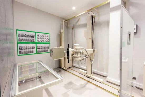
This condition is diagnosed based on X-ray scans. Computerized or magnetic resonance imaging is often performed to detail the detected abnormalities.
Conservative methods are used to treat a partially dislocated cervical vertebra. Manual repositioning of the displaced bone under anesthesia and subsequent immobilization is shown to restore the anatomy of the spinal column. However, in some cases, surgical intervention is indicated.
Types
Cervical vertebral subluxation can be:
- isolated;
- combined with other types of injuries.
Symptoms of cervical vertebral subluxation
The severity of the traumatic impact determines the symptoms of a cervical vertebra subluxation. In some cases, the patient may even lose consciousness. This condition is characterized by a forced turn of the head to the left or right and the impossibility of any movement in the cervical spine due to sharp pain in the occipital-cervical region. Even with gentle palpation of the cervical region, intense soreness is determined. Cervical muscles are in a spastic state, and swelling of soft tissue structures of the neck is detected. Painful sensations increase when trying to turn the head to one side or another. Dislocation of the second-third cervical vertebrae can create a mechanical obstacle in the path of the food clump, which causes difficulty in swallowing and neurotrophic tongue swelling. With subluxation of the sixth to seventh cervical vertebrae, painful sensations in the neck go to the shoulder region, and flatulence and discomfort may also appear in the chest area.
When nerve endings are compressed, the following clinical signs may occur:
- sleep disorders - insomnia at night and sleepiness during the day;
- convulsive muscle contractions (mainly tonic);
- painful sensations in the face, back, and arms (depending on the level of compression);
- the sensation of false tinnitus;
- paresthesia on the skin of the hand;
- muscle weakness in the arms.
Subluxation of the cervical vertebra in the newborn period is often hidden. However, as soon as the child tends to verticalize, the subluxation makes itself known by establishing an incorrect gait. Subsequently, such children complain of pain in the head, difficulty remembering, inability to concentrate, rapid fatigue, and frequent mood swings for no apparent reason.
Causes
Rotational dislocation of the atlas is common in childhood (in adults, it is a rarer condition). Causative factors can be:
- failures in physical education classes due to incorrectly turning the head to the side;
- sloppiness during active games or sports activities;
- application of external or internal force to the back of the head or neck.
A cervical vertebra subluxation can also be diagnosed immediately after birth. In this case, it is considered a birth trauma and is associated with a violation of the passage of the head through the birth canal. The predisposing factors are the pelvis's narrowness, bone deformities, and other obstacles. The ease of occurrence of cervical subluxations in the neonatal period is explained by the weakness of the ligamentous apparatus, as a result of which the vertebrae may be partially displaced from their normal anatomical position.
The following circumstances can be risk factors for cervical vertebrae subluxation, especially the second through seventh vertebrae:
- falling on the head in a state of slight flexion of the cervical region;
- unsuccessful diving with impact with the bottom of the water body;
- not following the tumbling technique;
- improper headstand;
- a blow to the back of the head when performing exercises with the head tilted downward;
- excessive flexion of the neck in a car accident, including previous overextension (so-called whiplash).
Diagnosis
Cervical subluxation can be detected based on anamnesis and clinical manifestations. However, the primary diagnostic tool is visualization using radial diagnostic methods, which allow the condition of bone structures and ligamentous apparatus to be assessed.
The primary method for detecting subluxation is radiography, initially performed in 2 central projections. However, lateral and direct scans cannot detect a displaced vertebra, so additional projections, including scans with functional loading (in flexion/extension of the cervical spine), are performed.
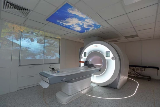
To determine the further patient management program, detailed examinations are also performed—CT or nuclear magnetic tomography. CT is more focused on evaluating anatomical formations with increased density, while MRI focuses on detailing soft tissue structures. A neurologist is consulted to evaluate the patient's nervous system. Echoencephalography allows us to determine the condition of the brain, and rheoencephalography helps to determine the nature of blood supply to the cerebral tissue.
Treatment of cervical vertebral subluxation
Conservative methods carry out treatment of cervical vertebral subluxation in most cases. At the same time, if there is a high probability of adverse neurological consequences and the injury is old, the issue is decided in favor of surgical intervention.
Conservative treatment
First aid consists of immobilizing the cervical region with special splints or improvised means. Subsequent conservative treatment involves fixating the cervical spine with an improvised cotton-gauze collar of the Shantz type and symptomatic therapy (pain relief, correction of cardiovascular disorders, etc.).
It is essential to correct the dislocation as early as possible. Swelling progresses over time, which can exacerbate the restoration of cervical anatomy. Redressing can be carried out by manual techniques or in stages using small weights. With one-stage redressing, a click is felt, which indicates that the displaced vertebra has taken the correct position. At the same time, the severity of the pain syndrome is reduced.
Surgical treatment
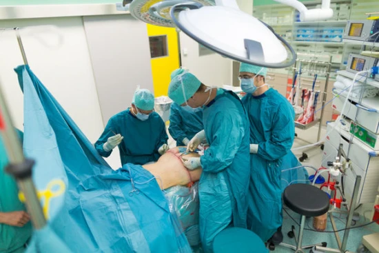
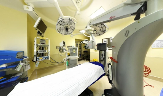
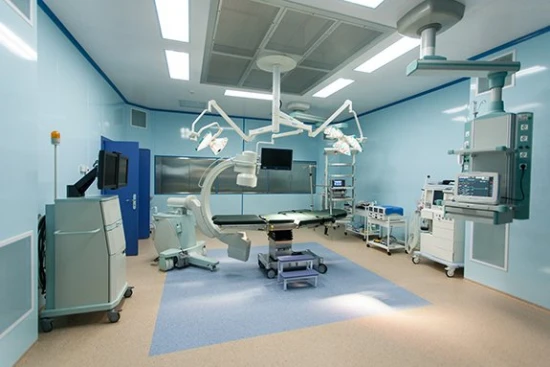
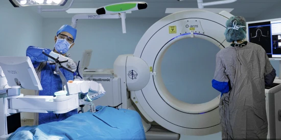
Surgery may be indicated for cervical spine instability if the injury is more than 1.5-2 months old. Atlantoaxial fixation surgery is also indicated when subluxation is combined with fracture of bony structures.
Prevention
Prevention of cervical vertebral subluxation consists of the following areas:
- rational and gentle management of labor;
- wearing seat belts before riding in a motor vehicle;
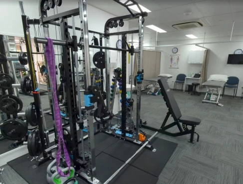
- strengthening the muscular corset of the spine;
- observing the correct technique of sports exercises;
- prevention of household injuries.
Rehabilitation
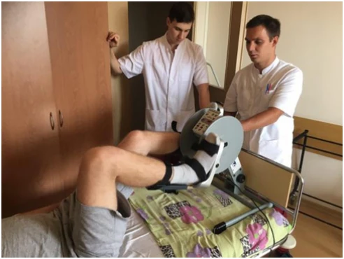
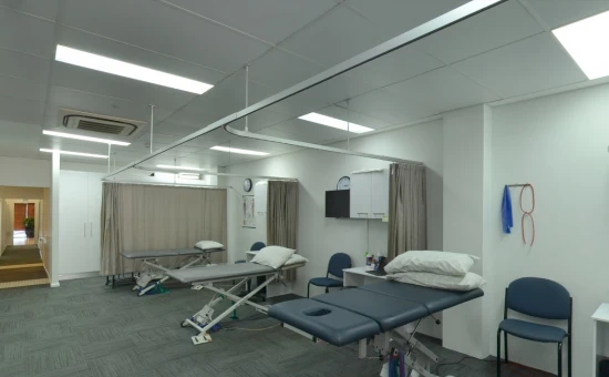
After a subluxation, including surgical treatment, comprehensive rehabilitation is indicated, which consists of the following areas:
- wearing a supportive corset (temporary measure; otherwise, secondary muscle atrophy may develop);
- cervical taping;
- various physical procedures (electrophoresis, ultrasound therapy, laser therapy, magnetotherapy);
- therapeutic massage.













-medium.webp)
-medium.webp)
-medium.webp)
-small.webp)


