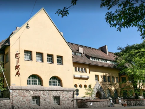
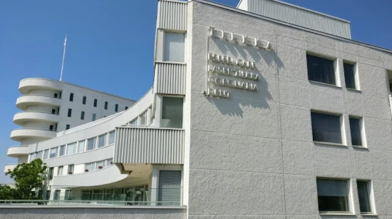
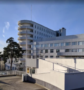
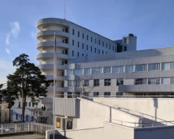
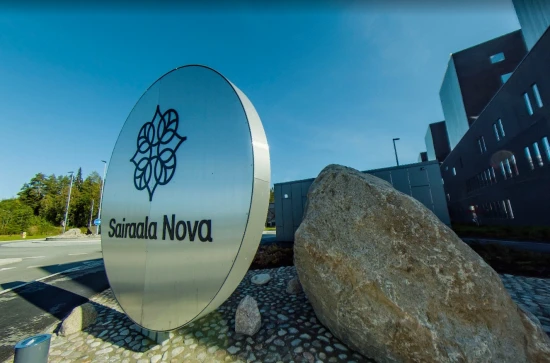
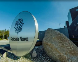
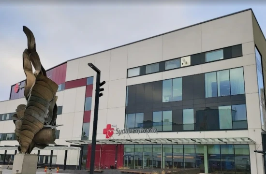
Thoracic aortic aneurysm (TAA) treatment in 1 Vascular surgery clinic in Finland
1 clinic specializing in Vascular surgery providing treatment of
Thoracic aortic aneurysm (TAA)
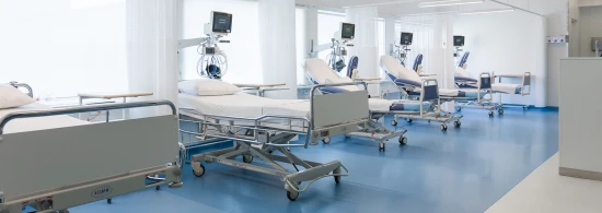
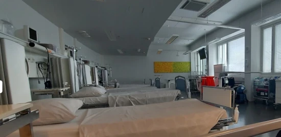
Thoracic aortic aneurysm (TAA) is a condition where the aorta's walls in the chest weaken and bulge, potentially leading to life-threatening complications if the aneurysm ruptures. Surveillance and, if necessary, surgical intervention are vital for managing TAA.
Read more...
disease in Finland.
Besides this clinic there are 6 Vascular surgery clinics in Finland.
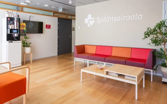
Such diseases are treated by Turku University Hospital (Tyks): Abdominal aortic aneurysm (AAA), Acute limb ischemia, Aortic dissection, Thoracic aortic aneurysm (TAA), Thoracoabdominal aortic aneurysm, and others.
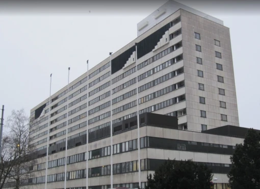


6 nearby similar clinics in Finland
Perhaps you should consider 6 more clinics we have found nearby basing on your Location, Disease filters applied.