Disease Types & Epidemiology
How common is the disease?
Cervical cancer starts in the cells lining the cervix, the lower part of the uterus that connects to the vagina. It is primarily caused by persistent infection with certain types of human papillomavirus, a common sexually transmitted virus. Despite being one of the most preventable cancers due to the availability of effective screening and vaccination programs, cervical cancer remains a significant public health concern, particularly in regions with limited resources.
Globally, cervical cancer is the fourth most common cancer among women, with an estimated 604,000 new cases and 342,000 deaths worldwide in 2020. In the United States, approximately 14,480 new cases of invasive cervical cancer are expected to be diagnosed in 2024, resulting in about 4,290 deaths. The incidence and mortality rates have significantly decreased over the past few decades due to widespread Pap test screening and HPV vaccination [SEER, 2024].
The two predominant types of cervical cancer are:
- Squamous cell carcinoma, which represents approximately 90% of cases and arises from the thin, flat cells covering the outer portion of the cervix that extends into the vagina.
- Adenocarcinoma accounts for around 10-20% of cervical cancers and develops from the glandular cells lining the cervical canal.
In addition, there are less common forms of cervical cancer, such as adenosquamous carcinomas and small-cell neuroendocrine tumors.
Causes & Risk Factors
What is the primary issue of cervical cancer?
The primary cause of cervical cancer is a persistent infection with certain high-risk strains of the human papillomavirus (HPV), particularly HPV 16 and 18, which account for around 70% of cervical cancer cases. Other factors that can increase the risk of developing this disease include engaging in sexual activity at a young age, having multiple sexual partners, smoking, having a weakened immune system (such as from HIV infection), using birth control pills for an extended period, giving birth to multiple children, and having limited access to screening and treatment services [Cancer.org 2023].
Clinical Manifestation & Symptoms
What signs should one anticipate while suspecting cervical cancer?
In the early stages, cervical cancer often does not cause any noticeable symptoms. However, as the disease progresses, individuals may start to experience certain signs, such as abnormal vaginal bleeding, including bleeding after sexual intercourse, between menstrual periods, or even after menopause. Additionally, some people may notice unusual vaginal discharge that can be watery, pink, or have an unpleasant odor. Furthermore, pelvic pain, which may occur during sexual activity or at other times, can also be a symptom of advancing cervical cancer. Experiencing pain during intercourse is another potential indicator that warrants medical attention.
Algorithm of diagnosis & staging
What evaluations do cervical cancer patients undergo to identify the best treatment strategy?
Cervical cancer staging helps determine the most appropriate treatment. Staging assesses the size, location, and potential spread of the cancer. The FIGO system is used to stage cervical cancer by evaluating tumor size, spread, and presence of distant metastases. Staging may involve several tests, such as:
- Examination under anesthesia is a detailed review of the cervix, vagina, uterus, bladder, and rectum, with biopsies of any abnormal areas to check for signs of cancer spread.
- Chest x-ray helps to detect any spread of cervical cancer to the lungs or chest cavity.
- An intravenous pyelogram is an x-ray of the urinary tract after injecting a special dye, used to identify any abnormal areas caused by the spread of cervical cancer.
Additional imaging techniques can be precious for disease staging and determining the most appropriate treatment approach:
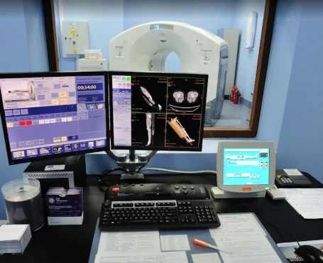
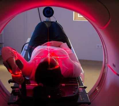
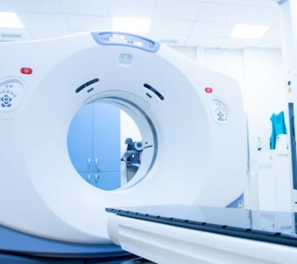
- Computed tomography (CT) scans provide three-dimensional X-ray images that can delineate the extent of the cancer. It may be used instead of other tests like chest X-rays and intravenous pyelograms for staging. These scans can also help detect if the cancer has spread to lymph nodes.
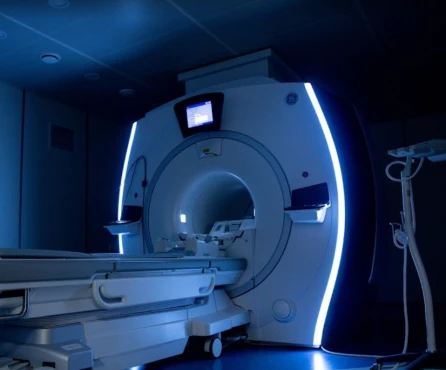
- Magnetic resonance imaging (MRI) scans leverage powerful magnetic fields and radio waves to generate highly detailed internal body images. MRI scanners can precisely determine the size and spread of the tumor with impressive accuracy.
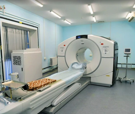
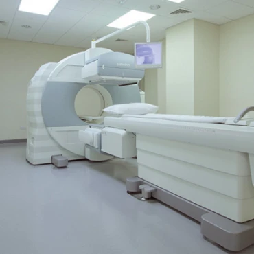
- Positron emission tomography (PET) scans utilize a radioactive tracer injected into the vein to highlight areas of abnormally high cellular activity, which can indicate the presence of cancer. PET scans are often combined with CT scans to provide comprehensive information about the cancer's characteristics and metastatic status.
Cervical cancer is staged using a combination of letter and number systems. The FIGO staging system utilizes Roman numerals I through IV, with lower stages generally indicating a better prognosis. The TNM system is also used alongside FIGO to evaluate three key factors:
- The size or extent of the primary tumor.
- Whether the cancer has spread to nearby lymph nodes.
- If the cancer has metastasized or spread to distant sites.
If a tumor biopsy is performed, it will be analyzed in the laboratory to determine the specific subtype of cervical cancer present.
Treatment routes
What is an appropriate treatment for different cervical cancer stages?
Treatment for cervical cancer is tailored based on the specific stage of the disease, the patient's overall health status, and their personal preferences. The primary therapeutic approaches encompass a range of options, including surgery, radiation therapy, chemotherapy, targeted therapies, and immunotherapies. These diverse treatment modalities allow clinicians to design the most appropriate and effective management plan for each patient.
For early-stage cervical cancer, there are several surgical options available. Conization, which involves removing a cone-shaped piece of tissue from the cervix, is often used for very early-stage disease and can effectively remove all cancer cells. For stage IA2 cervical cancer (3-7 mm invasion), a simple hysterectomy, where the uterus and cervix are removed, is typically the recommended procedure. This approach has an excellent prognosis, with about 93% of women surviving at least five years after treatment. For more advanced early-stage cervical cancer, such as stage IB (<4cm lesion) and IIA (>4 cm lesion), a radical hysterectomy may be performed. This more extensive surgery removes the uterus, cervix, part of the vagina, and surrounding tissues. Despite the more comprehensive nature of this procedure, it still has a high success rate, with approximately 85-90% of women living at least five years after this treatment.
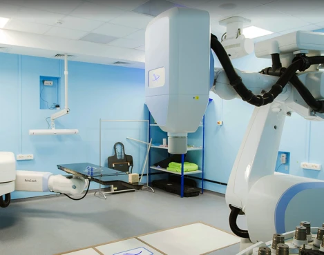
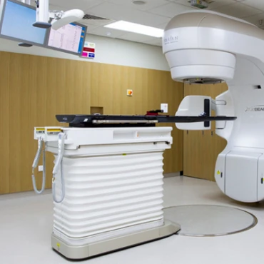
Radiation therapy plays a crucial role in the treatment of cervical cancer. External beam radiation therapy employs high-energy X-rays to target and destroy cancer cells. This approach is frequently combined with brachytherapy, a technique that involves placing radioactive material directly within or adjacent to the tumor. The combination of these two radiation modalities has shown impressive results, with around 80-85% of women achieving five-year survival rates.
Chemotherapy, particularly the use of the drug cisplatin, is often integrated with radiation therapy to enhance its effectiveness. This combined approach has demonstrated a response rate of around 80% in the early stages of cervical cancer.
For advanced-stage cervical cancer, there are a few fundamental treatment approaches. Chemotherapy options often include a combination of cisplatin and paclitaxel, which can yield partial response rates of around 40-60%. Alternatively, carboplatin may be used, especially for patients who cannot tolerate cisplatin. These chemotherapy drugs are frequently combined with other medications.
Additionally, radiation therapy plays a crucial role in managing advanced-stage cervical cancer. A combination of external beam radiation therapy (EBRT) and brachytherapy, which involves placing radioactive material directly in or near the tumor, can help control the spread of the cancer and provide symptom relief. For stage III cervical cancer patients treated with this radiation and chemotherapy approach, the five-year survival rate is approximately 50-60%.
For patients with refractory cervical cancer that is resistant to standard treatments, there are some promising targeted therapies and immunotherapies available. The targeted drug bevacizumab, which blocks the growth of new blood vessels that feed the cancer, has been shown to improve overall survival rates when combined with chemotherapy. This combination can increase median overall survival by around 3.7 months compared to chemotherapy alone.
Immunotherapy with the drug pembrolizumab has also emerged as an option, especially for cervical cancers that express the PD-L1 protein. Pembrolizumab works by "releasing the brakes" on the immune system, allowing it to attack the cancer cells more effectively. While the overall response rate is around 14.3%, some patients have experienced long-lasting remissions with this approach.
Prognosis & Follow-Up
How does cutting-edge science improve the lifespan and quality of life for those with the disease?
The prognosis for cervical cancer is highly dependent on the stage at which it is diagnosed. Patients diagnosed at earlier stages have significantly better 5-year survival rates, ranging from 80-93% for Stage I to just 15-16% for Stage IV. Fortunately, advancements in screening, HPV vaccination, and treatment approaches have dramatically improved outcomes for those battling this disease. Crucially, early detection through regular screening remains paramount to boosting survival and improving the outlook for cervical cancer patients.
Maintaining regular follow-up care is crucial for monitoring potential cancer recurrence and managing any side effects from treatment. The typical follow-up plan includes frequent physical exams, usually every 3 to 6 months for the first two years, followed by visits every 6 to 12 months for the next 3 to 5 years. Additionally, as the healthcare provider recommends, regular Pap tests are an essential part of the follow-up regimen. If needed, imaging tests such as CT scans or MRIs may also be used to monitor for any signs of the cancer returning.


